9FKO | pdb_00009fko

Arm segment of bacteriophage phi812


Experimental Data Snapshot

  • Method: X-RAY DIFFRACTION
  • Resolution: 1.80 Å
  • R-Value Free: 
    0.275 (Depositor), 0.275 (DCC) 
  • R-Value Work: 
    0.245 (Depositor), 0.245 (DCC) 
  • R-Value Observed: 
    0.247 (Depositor) 

wwPDB Validation 3D Report Full Report

Validation slider image for 9FKO

This is version 1.1 of the entry. See complete history

Literature

Conformational changes of the baseplate regulating tail contraction of Staphylococcus phage 812.

Binovsky, J.Siborova, M.Zlatohurska, M.Novacek, J.Bardy, P.Baska, R.Skubnik, K.Botka, T.Benesik, M.Pantucek, R.Tripsianes, K.Plevka, P.

(2026) EMBO J 

  • DOI: https://doi.org/10.1038/s44318-026-00834-9
  • Primary Citation Related Structures: 
    9EUJ, 9EUK, 9EUL, 9EUM, 9F04, 9F05, 9F06, 9FKO, 9TIC, 9TID, 9TIE, 9TIF, 9TIG, 9TIH, 9TII, 9TIJ, 9TIK, 9TIL, 9TIM, 9TIN, 9TIO, 9TIP, 9TIR, 9TIS, 9TIT, 9TIW

  • PubMed Abstract: 

    Phages with contractile tails employ elaborate mechanisms to penetrate bacterial cell walls and deliver their genomes into the host cytoplasm. Here, we used cryo-EM to show that the baseplate of phage 812, a member of the Kayvirus genus, which infects Gram-positive Staphylococcus strains, is formed of a core, wedge modules, and baseplate arms carrying receptor-binding proteins 1 and 2 and tripod complexes. Upon binding to a host cell, the receptor-binding proteins of phage 812 baseplate reorient and undergo conformational changes. The changes to the tripod complexes trigger the release of the central spike and weld proteins, which expose peptidoglycan-degrading domains of the hub proteins. Changes in the positions of baseplate arms are transmitted through wedge modules to tail sheath initiator proteins. The ring of the tail sheath initiator proteins expands and triggers the contraction of the tail sheath, which shortens to 50% and pushes the tail tube 10-30 nm into the bacterial cytoplasm. Homologous molecular mechanisms are probably shared by phages of the Herelleviridae family with contractile tails to infect Gram-positive bacteria.


  • Organizational Affiliation
    • Central European Institute of Technology, Brno, Czech Republic.

Macromolecule Content 

  • Total Structure Weight: 116.66 kDa 
  • Atom Count: 8,239 
  • Modeled Residue Count: 1,051 
  • Deposited Residue Count: 1,051 
  • Unique protein chains: 2

Macromolecules

Find similar proteins by:|  3D Structure
Entity ID: 1
MoleculeChains  Sequence LengthOrganismDetailsImage
Baseplate protein
A, B, C, D, E
A, B, C, D, E, F
174Staphylococcus phage 812Mutation(s): 0 
Gene Names: 812_119812_121812a_121812F1_121K1/420_121K1_121
UniProt
Find proteins for Q4Z9Z7 (Staphylococcus phage G1)
Explore Q4Z9Z7 
Go to UniProtKB:  Q4Z9Z7
Entity Groups
Sequence Clusters30% Identity50% Identity70% Identity90% Identity95% Identity100% Identity
UniProt GroupQ4Z9Z7
Sequence Annotations
Expand
Reference Sequence
Find similar proteins by:  Sequence   |   3D Structure  
Entity ID: 2
MoleculeChains  Sequence LengthOrganismDetailsImage
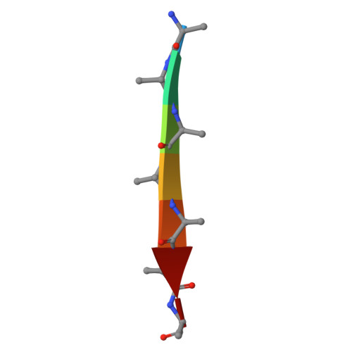
Unknown peptide7Escherichia coliMutation(s): 0 

Experimental Data & Validation

Experimental Data

  • Method: X-RAY DIFFRACTION
  • Resolution: 1.80 Å
  • R-Value Free:  0.275 (Depositor), 0.275 (DCC) 
  • R-Value Work:  0.245 (Depositor), 0.245 (DCC) 
  • R-Value Observed: 0.247 (Depositor) 
Space Group: P 1 21 1
Unit Cell:
Length ( Å )Angle ( ˚ )
a = 92.93α = 90
b = 75.14β = 109.87
c = 94.45γ = 90
Software Package:
Software NamePurpose
PHENIXrefinement
PHENIXrefinement
XDSdata reduction
XDSdata scaling
SHELXDphasing

Structure Validation

View Full Validation Report


Entry History 

& Funding Information

Deposition Data


Funding OrganizationLocationGrant Number
European Research Council (ERC)European Union101043452
Other governmentCzech RepublicLX22NP05103

Revision History  (Full details and data files)

  • Version 1.0: 2025-06-18
    Type: Initial release
  • Version 1.1: 2026-07-08
    Changes: Database references