9FKD | pdb_00009fkd

Progesterone-bound DB3 Fab in complex with computationally designed DBPro1156_2 protein binder


Experimental Data Snapshot

  • Method: ELECTRON MICROSCOPY
  • Resolution: 3.30 Å
  • Aggregation State: PARTICLE 
  • Reconstruction Method: SINGLE PARTICLE 

wwPDB Validation 3D Report Full Report

Validation slider image for 9FKD

This is version 1.4 of the entry. See complete history

Literature

Targeting protein-ligand neosurfaces with a generalizable deep learning tool.

Marchand, A.Buckley, S.Schneuing, A.Pacesa, M.Elia, M.Gainza, P.Elizarova, E.Neeser, R.M.Lee, P.W.Reymond, L.Miao, Y.Scheller, L.Georgeon, S.Schmidt, J.Schwaller, P.Maerkl, S.J.Bronstein, M.Correia, B.E.

(2025) Nature 639: 522-531

  • DOI: https://doi.org/10.1038/s41586-024-08435-4
  • Primary Citation Related Structures: 
    8S1X, 9FKD

  • PubMed Abstract: 

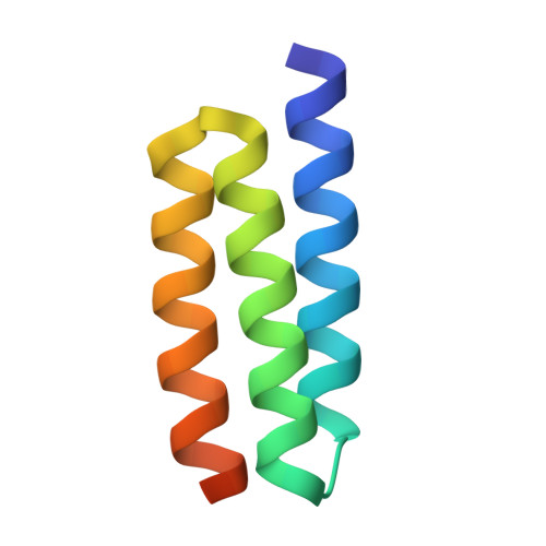
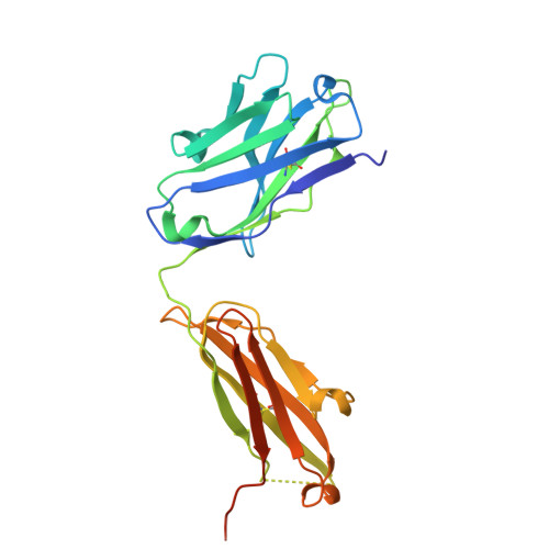
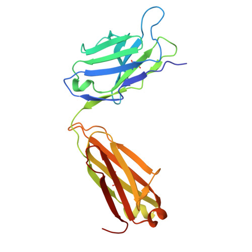
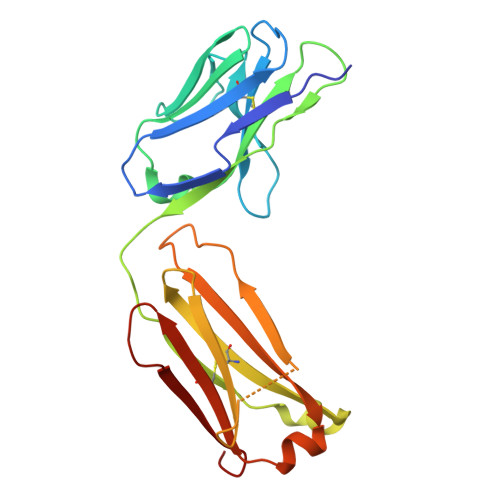
    Molecular recognition events between proteins drive biological processes in living systems 1 . However, higher levels of mechanistic regulation have emerged, in which protein-protein interactions are conditioned to small molecules 2-5 . Despite recent advances, computational tools for the design of new chemically induced protein interactions have remained a challenging task for the field 6,7 . Here we present a computational strategy for the design of proteins that target neosurfaces, that is, surfaces arising from protein-ligand complexes. To develop this strategy, we leveraged a geometric deep learning approach based on learned molecular surface representations 8,9 and experimentally validated binders against three drug-bound protein complexes: Bcl2-venetoclax, DB3-progesterone and PDF1-actinonin. All binders demonstrated high affinities and accurate specificities, as assessed by mutational and structural characterization. Remarkably, surface fingerprints previously trained only on proteins could be applied to neosurfaces induced by interactions with small molecules, providing a powerful demonstration of generalizability that is uncommon in other deep learning approaches. We anticipate that such designed chemically induced protein interactions will have the potential to expand the sensing repertoire and the assembly of new synthetic pathways in engineered cells for innovative drug-controlled cell-based therapies 10 .


  • Organizational Affiliation
    • Laboratory of Protein Design and Immunoengineering, Institute of Bioengineering, Ecole polytechnique fédérale de Lausanne, Lausanne, Switzerland.

Macromolecule Content 

  • Total Structure Weight: 108.13 kDa 
  • Atom Count: 7,193 
  • Modeled Residue Count: 933 
  • Deposited Residue Count: 983 
  • Unique protein chains: 5

Macromolecules

Find similar proteins by:|  3D Structure
Entity ID: 1
MoleculeChains  Sequence LengthOrganismDetailsImage
De novo designed DBPro1156_2 binderA [auth B]70synthetic constructMutation(s): 0 
Find similar proteins by:|  3D Structure
Entity ID: 2
MoleculeChains  Sequence LengthOrganismDetailsImage
DB3 Fab Heavy chainB [auth H]239synthetic constructMutation(s): 0 
Find similar proteins by:|  3D Structure
Entity ID: 3
MoleculeChains  Sequence LengthOrganismDetailsImage
DB3 Fab Light ChainC [auth L]222synthetic constructMutation(s): 0 
Find similar proteins by:|  3D Structure
Entity ID: 4
MoleculeChains  Sequence LengthOrganismDetailsImage
Anti-kappa Fab Light ChainD [auth K]217synthetic constructMutation(s): 0 
Find similar proteins by:|  3D Structure
Entity ID: 5
MoleculeChains  Sequence LengthOrganismDetailsImage
Anti-kappa Fab Heavy ChainE [auth I]235synthetic constructMutation(s): 0 

Small Molecules

Ligands 1 Unique
IDChains Name / Formula / InChI Key2D Diagram3D Interactions
STR
(Subject of Investigation/LOI)

Query on STR


Download:Ideal Coordinates CCD File
F [auth H]PROGESTERONE
C21 H30 O2
RJKFOVLPORLFTN-LEKSSAKUSA-N

Experimental Data & Validation

Experimental Data

  • Method: ELECTRON MICROSCOPY
  • Resolution: 3.30 Å
  • Aggregation State: PARTICLE 
  • Reconstruction Method: SINGLE PARTICLE 
EM Software:
TaskSoftware PackageVersion
MODEL REFINEMENTPHENIXdev_5316
RECONSTRUCTIONcryoSPARC4.4.1

Structure Validation

View Full Validation Report


Entry History 

& Funding Information

Deposition Data


Funding OrganizationLocationGrant Number
Swiss National Science FoundationSwitzerland310030_197724

Revision History  (Full details and data files)

  • Version 1.0: 2024-10-30
    Type: Initial release
  • Version 1.1: 2024-11-20
    Changes: Data collection
  • Version 1.2: 2025-01-15
    Changes: Data collection, Database references
  • Version 1.3: 2025-01-29
    Changes: Data collection, Database references
  • Version 1.4: 2025-03-26
    Changes: Data collection, Database references