9FD0 | pdb_00009fd0

Structure of the Saccharomyces cerevisiae Pmt4-MIR domain with bound ligands


Experimental Data Snapshot

  • Method: X-RAY DIFFRACTION
  • Resolution: 1.35 Å
  • R-Value Free: 
    0.191 (Depositor), 0.191 (DCC) 
  • R-Value Work: 
    0.152 (Depositor), 0.153 (DCC) 
  • R-Value Observed: 
    0.154 (Depositor) 

Starting Model: experimental
View more details

wwPDB Validation   3D Report Full Report


This is version 1.1 of the entry. See complete history


Literature

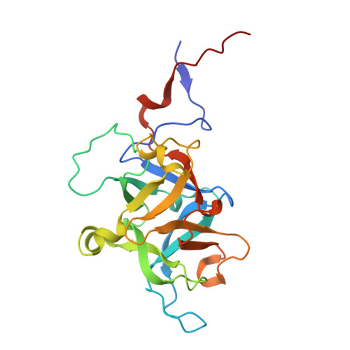
Structural characterisation of the fungal Pmt4 homodimer.

McDowell, M.A.Wild, K.Fiorentino, F.Bausewein, D.Metschies, A.Chiapparino, A.Hackmann, Y.Bilsing, F.L.Brenske, D.Mortensen, S.Wu, D.Robinson, C.V.Strahl, S.Sinning, I.

(2025) Nat Commun 16: 11134-11134

  • DOI: https://doi.org/10.1038/s41467-025-67412-1
  • Primary Citation of Related Structures:  
    9FD0, 9FD1, 9I5K, 9I5L

  • PubMed Abstract: 

    Protein O-mannosyltransferases (PMTs) are conserved endoplasmic reticulum membrane-embedded enzymes responsible for the transfer of mannose from dolichol phosphate-mannose (Dol-P-Man) to serine/threonine-rich protein substrates or unfolded proteins. PMTs from three subfamilies form obligate dimers with different substrate specificities and require the concerted action of their transmembrane domains (TMDs) and a luminal MIR domain for catalysis. Here, we present structures, native mass spectrometry, and structure-based mutagenesis of the fungal Pmt4 homodimer. The core fold of the TMDs and MIR domain is conserved with the Pmt1-Pmt2 heterodimer, indicating a shared catalytic mechanism. Distinct from Pmt4, the MIR domain interacts in cis with the TMDs of the same subunit and has a β-hairpin insertion required for O-mannosylation of substrates. We further identify a cytosolic binding site for substrate Dol-P-Man within the Pmt4 TMDs, which is conserved amongst PMTs and important for in vivo activity. Thus, we provide a framework to understand the substrate specificity and regulation of the Pmt4 homodimer.


  • Organizational Affiliation
    • Heidelberg University Biochemistry Centre (BZH), Heidelberg, Germany. melanie.mcdowell@biophys.mpg.de.

Macromolecules
Find similar proteins by:  (by identity cutoff)  |  3D Structure
Entity ID: 1
MoleculeChains Sequence LengthOrganismDetailsImage
Dolichyl-phosphate-mannose--protein mannosyltransferase 4222Saccharomyces cerevisiaeMutation(s): 0 
Gene Names: PMT4YJR143CJ2176
EC: 2.4.1.109
UniProt
Find proteins for P46971 (Saccharomyces cerevisiae (strain ATCC 204508 / S288c))
Explore P46971 
Go to UniProtKB:  P46971
Entity Groups  
Sequence Clusters30% Identity50% Identity70% Identity90% Identity95% Identity100% Identity
UniProt GroupP46971
Sequence Annotations
Expand
  • Reference Sequence
Experimental Data & Validation

Experimental Data

  • Method: X-RAY DIFFRACTION
  • Resolution: 1.35 Å
  • R-Value Free:  0.191 (Depositor), 0.191 (DCC) 
  • R-Value Work:  0.152 (Depositor), 0.153 (DCC) 
  • R-Value Observed: 0.154 (Depositor) 
Space Group: P 21 21 21
Unit Cell:
Length ( Å )Angle ( ˚ )
a = 48.593α = 90
b = 56.767β = 90
c = 78.216γ = 90
Software Package:
Software NamePurpose
PHENIXrefinement
XDSdata reduction
Aimlessdata scaling
PHENIXphasing

Structure Validation

View Full Validation Report


Entry History & Funding Information

Deposition Data


Funding OrganizationLocationGrant Number
German Research Foundation (DFG)Germany--

Revision History  (Full details and data files)

  • Version 1.0: 2025-11-26
    Type: Initial release
  • Version 1.1: 2026-01-21
    Changes: Database references, Structure summary