9F0O | pdb_00009f0o

The molecular basis and modulation of lamin-specific chromatin interaction


Experimental Data Snapshot

  • Method: ELECTRON MICROSCOPY
  • Resolution: 2.30 Å
  • Aggregation State: PARTICLE 
  • Reconstruction Method: SINGLE PARTICLE 

wwPDB Validation   3D Report Full Report


This is version 1.1 of the entry. See complete history


Literature

The molecular basis of lamin-specific chromatin interactions.

Wang, B.Kronenberg-Tenga, R.Rosti, V.Di Patrizio Soldateschi, E.Luo, Q.Iannacchero, U.M.Pinet, L.Eibauer, M.Boujemaa-Paterski, R.Schuler, B.Lanzuolo, C.Medalia, O.

(2025) Nat Struct Mol Biol 32: 1999-2011

  • DOI: https://doi.org/10.1038/s41594-025-01622-5
  • Primary Citation of Related Structures:  
    9F0O

  • PubMed Abstract: 

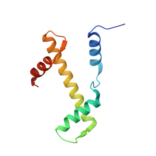
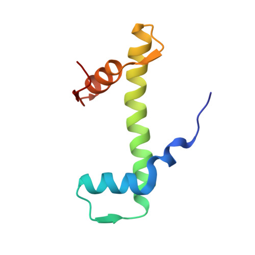
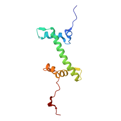
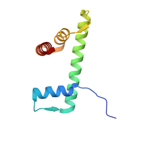
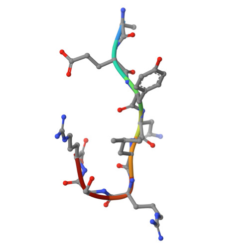
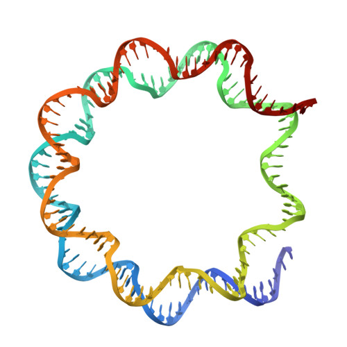
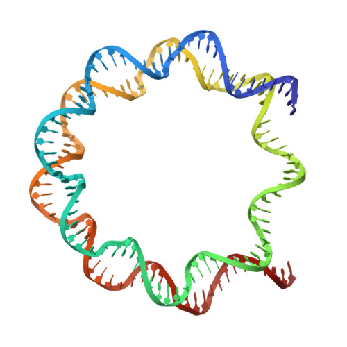
    In the cell nucleus, chromatin is anchored to the nuclear lamina, a network of lamin filaments and binding proteins that underly the inner nuclear membrane. The nuclear lamina is involved in chromatin organization through the interaction of lamina-associated domains within the densely packed heterochromatin regions. Using cryo-focused ion beam milling in conjunction with cryo-electron tomography, we analyzed the distribution of nucleosomes at the lamin-chromatin interface at the nanometer scale. Depletion of lamins A and C reduced nucleosome concentration at the nuclear periphery, while B-type lamin depletion contributed to nucleosome density in proximity to the lamina but not further away. We then investigated whether specific lamins can mediate direct interactions with chromatin. Using cryo-electron microscopy, we identified a specific binding motif of the lamin A tail domain that interacts with nucleosomes, distinguishing it from the other lamin isoforms. Furthermore, we examined chromatin structure dynamics using a genome-wide analysis that revealed lamin-dependent macroscopic-scale alterations in gene expression and chromatin remodeling. Our findings provide detailed insights into the dynamic and structural interplay between lamin isoforms and chromatin, molecular interactions that shape chromatin architecture and epigenetic regulation.


  • Organizational Affiliation
    • Department of Biochemistry, University of Zurich, Zurich, Switzerland.

Macromolecules

Find similar proteins by:  (by identity cutoff)  |  3D Structure
Entity ID: 1
MoleculeChains Sequence LengthOrganismDetailsImage
Histone H3.2
A, E
98Xenopus laevisMutation(s): 0 
UniProt
Find proteins for P84233 (Xenopus laevis)
Explore P84233 
Go to UniProtKB:  P84233
Entity Groups  
Sequence Clusters30% Identity50% Identity70% Identity90% Identity95% Identity100% Identity
UniProt GroupP84233
Sequence Annotations
Expand
  • Reference Sequence
Find similar proteins by:  (by identity cutoff)  |  3D Structure
Entity ID: 2
MoleculeChains Sequence LengthOrganismDetailsImage
Histone H4
B, F
83Xenopus laevisMutation(s): 0 
UniProt
Find proteins for P62799 (Xenopus laevis)
Explore P62799 
Go to UniProtKB:  P62799
Entity Groups  
Sequence Clusters30% Identity50% Identity70% Identity90% Identity95% Identity100% Identity
UniProt GroupP62799
Sequence Annotations
Expand
  • Reference Sequence
Find similar proteins by:  (by identity cutoff)  |  3D Structure
Entity ID: 3
MoleculeChains Sequence LengthOrganismDetailsImage
Histone H2A type 1
C, G
110Xenopus laevisMutation(s): 0 
UniProt
Find proteins for P06897 (Xenopus laevis)
Explore P06897 
Go to UniProtKB:  P06897
Entity Groups  
Sequence Clusters30% Identity50% Identity70% Identity90% Identity95% Identity100% Identity
UniProt GroupP06897
Sequence Annotations
Expand
  • Reference Sequence
Find similar proteins by:  (by identity cutoff)  |  3D Structure
Entity ID: 4
MoleculeChains Sequence LengthOrganismDetailsImage
Histone H2B 1.1
D, H
97Xenopus laevisMutation(s): 0 
UniProt
Find proteins for P02281 (Xenopus laevis)
Explore P02281 
Go to UniProtKB:  P02281
Entity Groups  
Sequence Clusters30% Identity50% Identity70% Identity90% Identity95% Identity100% Identity
UniProt GroupP02281
Sequence Annotations
Expand
  • Reference Sequence

Find similar proteins by:  Sequence   |   3D Structure  

Entity ID: 7
MoleculeChains Sequence LengthOrganismDetailsImage
laminA_peptide
K, L
8Homo sapiensMutation(s): 0 
Sequence Annotations
Expand
  • Reference Sequence
Find similar nucleic acids by:  (by identity cutoff)  |  3D Structure
Entity ID: 5
MoleculeChains LengthOrganismImage
601 wisdom DNA147Homo sapiens
Sequence Annotations
Expand
  • Reference Sequence
Find similar nucleic acids by:  (by identity cutoff)  |  3D Structure
Entity ID: 6
MoleculeChains LengthOrganismImage
601 wisdom DNA147Homo sapiens
Sequence Annotations
Expand
  • Reference Sequence
Experimental Data & Validation

Experimental Data

  • Method: ELECTRON MICROSCOPY
  • Resolution: 2.30 Å
  • Aggregation State: PARTICLE 
  • Reconstruction Method: SINGLE PARTICLE 
EM Software:
TaskSoftware PackageVersion
MODEL REFINEMENTPHENIX

Structure Validation

View Full Validation Report


Entry History & Funding Information

Deposition Data


Funding OrganizationLocationGrant Number
Swiss National Science FoundationSwitzerlandSNSF 391 310030_207453

Revision History  (Full details and data files)

  • Version 1.0: 2025-08-13
    Type: Initial release
  • Version 1.1: 2025-10-22
    Changes: Data collection, Database references