


Redesigning surface charge to control substrate morphology preference of a PET-hydrolase
Oliveira-Pessoa, L., Lichtenstein, B.R.To be published.
Experimental Data Snapshot
Starting Model: in silico
View more details
wwPDB Validation 3D Report Full Report

Entity ID: 1 | |||||
|---|---|---|---|---|---|
| Molecule | Chains | Sequence Length | Organism | Details | Image |
| cutinase | 269 | Saccharopolyspora flava | Mutation(s): 23 Gene Names: SAMN05660874_00127 EC: 3.1.1.74 | 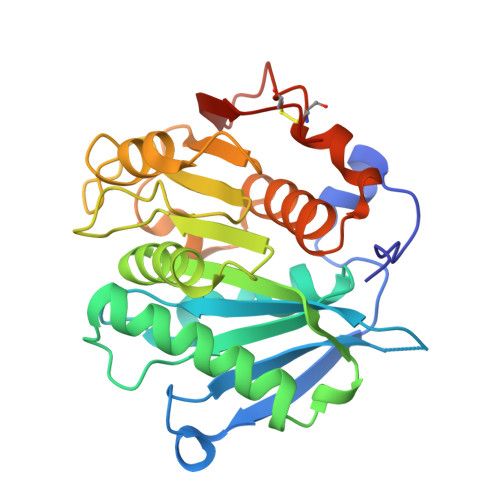 | |
UniProt | |||||
Find proteins for A0A1I6NU60 (Saccharopolyspora flava) Explore A0A1I6NU60 Go to UniProtKB: A0A1I6NU60 | |||||
Entity Groups | |||||
| Sequence Clusters | 30% Identity50% Identity70% Identity90% Identity95% Identity100% Identity | ||||
| UniProt Group | A0A1I6NU60 | ||||
Sequence AnnotationsExpand | |||||
Reference Sequence | |||||
| Length ( Å ) | Angle ( ˚ ) |
|---|---|
| a = 46.084 | α = 90 |
| b = 41.316 | β = 99.815 |
| c = 124.088 | γ = 90 |
| Software Name | Purpose |
|---|---|
| REFMAC | refinement |
| autoPROC | data reduction |
| STARANISO | data scaling |
| MOLREP | phasing |
| Funding Organization | Location | Grant Number |
|---|---|---|
| UK Research and Innovation (UKRI) | United Kingdom | Research England |


