9EVL | pdb_00009evl

Crystal Structure of human Collagen Hydroxylysine Galactosyltransferase GLT25D1/COLGALT1: Hg2+ soak for experimental phasing


Experimental Data Snapshot

  • Method: X-RAY DIFFRACTION
  • Resolution: 2.80 Å
  • R-Value Free: 
    0.239 (Depositor), 0.252 (DCC) 
  • R-Value Work: 
    0.202 (Depositor), 0.212 (DCC) 
  • R-Value Observed: 
    0.204 (Depositor) 

wwPDB Validation   3D Report Full Report


This is version 1.1 of the entry. See complete history


Literature

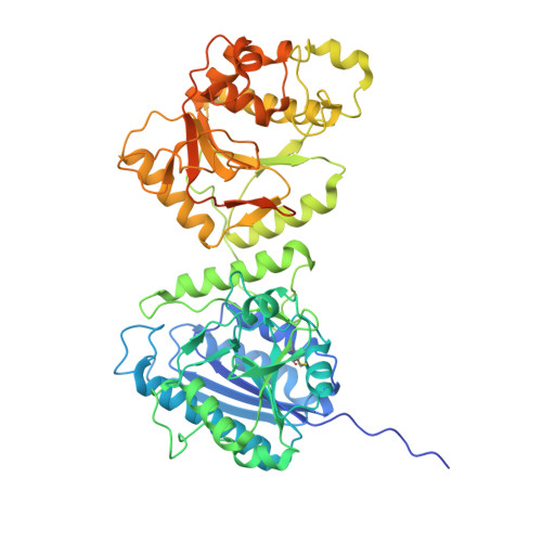
Molecular structure and enzymatic mechanism of the human collagen hydroxylysine galactosyltransferase GLT25D1/COLGALT1.

De Marco, M.Rai, S.R.Scietti, L.Mattoteia, D.Liberi, S.Moroni, E.Pinnola, A.Vetrano, A.Iacobucci, C.Santambrogio, C.Colombo, G.Forneris, F.

(2025) Nat Commun 16: 3624-3624

  • DOI: https://doi.org/10.1038/s41467-025-59017-5
  • Primary Citation of Related Structures:  
    9EVJ, 9EVK, 9EVL

  • PubMed Abstract: 

    During collagen biosynthesis, lysine residues undergo extensive post-translational modifications through the alternate action of two distinct metal ion-dependent enzyme families (i.e., LH/PLODs and GLT25D/COLGALT), ultimately producing the highly conserved α-(1,2)-glucosyl-β-(1,O)-galactosyl-5-hydroxylysine pattern. Malfunctions in these enzymes are linked to developmental pathologies and extracellular matrix alterations associated to enhanced aggressiveness of solid tumors. Here, we characterized human GLT25D1/COLGALT1, revealing an elongated head-to-head homodimeric assembly. Each monomer encompasses two domains (named GT1 and GT2), both unexpectedly capable of binding metal ion cofactors and UDP-α-galactose donor substrates, resulting in four candidate catalytic sites per dimer. We identify the catalytic site in GT2, featuring an unusual Glu-Asp-Asp motif critical for Mn 2+ binding, ruling out direct catalytic roles for the GT1 domain, but showing that in this domain the unexpectedly bound Ca 2+ and UDP-α-galactose cofactors are critical for folding stability. Dimerization, albeit not essential for GLT25D1/COLGALT1 activity, provides a critical molecular contact site for multi-enzyme assembly interactions with partner multifunctional LH/PLOD lysyl hydroxylase-glycosyltransferase enzymes.


  • Organizational Affiliation
    • The Armenise-Harvard Laboratory of Structural Biology, Dept. Biology and Biotechnology, University of Pavia, Via Ferrata 9A, 27100, Pavia, Italy.

Macromolecules
Find similar proteins by:  (by identity cutoff)  |  3D Structure
Entity ID: 1
MoleculeChains Sequence LengthOrganismDetailsImage
Procollagen galactosyltransferase 1
A, B
594Homo sapiensMutation(s): 0 
Gene Names: COLGALT1GLT25D1PSEC0241
EC: 2.4.1.50
UniProt & NIH Common Fund Data Resources
Find proteins for Q8NBJ5 (Homo sapiens)
Explore Q8NBJ5 
Go to UniProtKB:  Q8NBJ5
PHAROS:  Q8NBJ5
GTEx:  ENSG00000130309 
Entity Groups  
Sequence Clusters30% Identity50% Identity70% Identity90% Identity95% Identity100% Identity
UniProt GroupQ8NBJ5
Sequence Annotations
Expand
  • Reference Sequence
Small Molecules
Ligands 5 Unique
IDChains Name / Formula / InChI Key2D Diagram3D Interactions
GDU (Subject of Investigation/LOI)
Query on GDU

Download Ideal Coordinates CCD File 
L [auth A],
W [auth B]
GALACTOSE-URIDINE-5'-DIPHOSPHATE
C15 H24 N2 O17 P2
HSCJRCZFDFQWRP-ABVWGUQPSA-N
HG
Query on HG

Download Ideal Coordinates CCD File 
C [auth A]
D [auth A]
E [auth A]
F [auth A]
G [auth A]
C [auth A],
D [auth A],
E [auth A],
F [auth A],
G [auth A],
P [auth B],
Q [auth B],
R [auth B],
S [auth B]
MERCURY (II) ION
Hg
BQPIGGFYSBELGY-UHFFFAOYSA-N
MES
Query on MES

Download Ideal Coordinates CCD File 
M [auth A],
N [auth A]
2-(N-MORPHOLINO)-ETHANESULFONIC ACID
C6 H13 N O4 S
SXGZJKUKBWWHRA-UHFFFAOYSA-N
CA (Subject of Investigation/LOI)
Query on CA

Download Ideal Coordinates CCD File 
O [auth A],
X [auth B]
CALCIUM ION
Ca
BHPQYMZQTOCNFJ-UHFFFAOYSA-N
CL
Query on CL

Download Ideal Coordinates CCD File 
H [auth A]
I [auth A]
J [auth A]
K [auth A]
T [auth B]
H [auth A],
I [auth A],
J [auth A],
K [auth A],
T [auth B],
U [auth B],
V [auth B]
CHLORIDE ION
Cl
VEXZGXHMUGYJMC-UHFFFAOYSA-M
Experimental Data & Validation

Experimental Data

  • Method: X-RAY DIFFRACTION
  • Resolution: 2.80 Å
  • R-Value Free:  0.239 (Depositor), 0.252 (DCC) 
  • R-Value Work:  0.202 (Depositor), 0.212 (DCC) 
  • R-Value Observed: 0.204 (Depositor) 
Space Group: I 2 3
Unit Cell:
Length ( Å )Angle ( ˚ )
a = 220.187α = 90
b = 220.187β = 90
c = 220.187γ = 90
Software Package:
Software NamePurpose
PHENIXrefinement
XDSdata reduction
Aimlessdata scaling
HKL2Mapphasing

Structure Validation

View Full Validation Report


Entry History & Funding Information

Deposition Data


Funding OrganizationLocationGrant Number
Italian Association for Cancer ResearchItalyMFAG 20075
Italian Association for Cancer ResearchItalyBRIDGE 27004
The Giovanni Armenise-Harvard FoundationUnited StatesCDA 2013
Mizutani Foundation for GlycoscienceJapan200039
Other privateUnited States--

Revision History  (Full details and data files)

  • Version 1.0: 2025-04-09
    Type: Initial release
  • Version 1.1: 2025-04-23
    Changes: Database references, Derived calculations