9EUV | pdb_00009euv

Lymphostatin, conformation 1 (composite structure)


Experimental Data Snapshot

  • Method: ELECTRON MICROSCOPY
  • Resolution: 3.00 Å
  • Aggregation State: PARTICLE 
  • Reconstruction Method: SINGLE PARTICLE 

Starting Model: experimental
View more details

wwPDB Validation   3D Report Full Report


This is version 1.2 of the entry. See complete history


Literature

Structure of lymphostatin, a large multi-functional virulence factor of pathogenic Escherichia coli.

Griessmann, M.Rasmussen, T.Flegler, V.J.Kraft, C.Schneider, R.Hateley, M.Spantzel, L.Stevens, M.P.Borsch, M.Bottcher, B.

(2025) Nat Commun 16: 5389-5389

  • DOI: https://doi.org/10.1038/s41467-025-60995-9
  • Primary Citation of Related Structures:  
    9EUV, 9EUW, 9QB8, 9QBB, 9QHH

  • PubMed Abstract: 

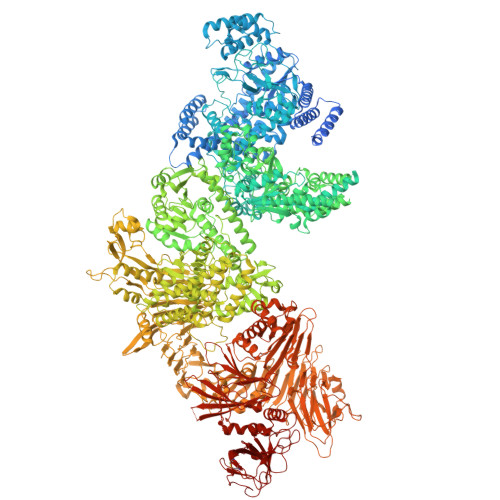
    Lymphostatin is a key virulence factor of enteropathogenic and enterohaemorrhagic Escherichia coli, playing roles in bacterial colonisation of the gut and in the inhibition of lymphocyte proliferation and proinflammatory responses. The protein's glycosyltransferase and cysteine protease motifs are required for activity against lymphocytes, but high-resolution structural information has proven elusive. Here, we describe the structure of lymphostatin from enteropathogenic E. coli O127:H6, determined by electron cryo-microscopy at different pH values. We observe three conformations of a highly complex molecule with two glycosyltransferase domains, one PaToxP-like protease domain, an ADP-ribosyltransferase domain, a vertex domain and a delivery domain. Long linkers hold these domains together and occlude the catalytic sites of the N-terminal glycosyltransferase and protease domains. Lymphostatin binds to bovine T-lymphocytes and HEK-293T cells, forming clusters at the plasma membrane that are internalized. With six distinct domains, lymphostatin can be regarded as a multitool of pathogenic Escherichia coli, enabling complex interactions with host cells.


  • Organizational Affiliation
    • University of Würzburg, Rudolf Virchow Center and Biocenter, Würzburg, Germany.

Macromolecules
Find similar proteins by:  (by identity cutoff)  |  3D Structure
Entity ID: 1
MoleculeChains Sequence LengthOrganismDetailsImage
Efa1/LifA protein3,229Escherichia coli O127:H6 str. E2348/69Mutation(s): 0 
Gene Names: E2348C_3234
UniProt
Find proteins for B7UI23 (Escherichia coli O127:H6 (strain E2348/69 / EPEC))
Explore B7UI23 
Go to UniProtKB:  B7UI23
Entity Groups  
Sequence Clusters30% Identity50% Identity70% Identity90% Identity95% Identity100% Identity
UniProt GroupB7UI23
Sequence Annotations
Expand
  • Reference Sequence
Experimental Data & Validation

Experimental Data

  • Method: ELECTRON MICROSCOPY
  • Resolution: 3.00 Å
  • Aggregation State: PARTICLE 
  • Reconstruction Method: SINGLE PARTICLE 
EM Software:
TaskSoftware PackageVersion
RECONSTRUCTIONRELION5.0
MODEL REFINEMENTPHENIX1.20.1

Structure Validation

View Full Validation Report


Entry History & Funding Information

Deposition Data


Funding OrganizationLocationGrant Number
German Research Foundation (DFG)Germany359471283
German Research Foundation (DFG)Germany456578072
German Research Foundation (DFG)Germany525040890
German Research Foundation (DFG)Germany428774170
German Research Foundation (DFG)GermanyINST 93/903-1
German Research Foundation (DFG)GermanyINST 93/1042-1
German Research Foundation (DFG)GermanyINST 93/1143-1
German Research Foundation (DFG)GermanyBO1150/18-1

Revision History  (Full details and data files)

  • Version 1.0: 2025-04-09
    Type: Initial release
  • Version 1.1: 2025-07-16
    Changes: Data collection, Database references
  • Version 1.2: 2025-07-23
    Changes: Data collection