9ETI | pdb_00009eti

Complex structure of IL-36R D1-D2 domain with 36R-D481 and BI655130 Fab


Experimental Data Snapshot

  • Method: X-RAY DIFFRACTION
  • Resolution: 2.40 Å
  • R-Value Free: 
    0.216 (Depositor), 0.205 (DCC) 
  • R-Value Work: 
    0.189 (Depositor), 0.177 (DCC) 
  • R-Value Observed: 
    0.190 (Depositor) 

Starting Model: experimental
View more details

wwPDB Validation   3D Report Full Report


Ligand Structure Quality Assessment 


This is version 1.0 of the entry. See complete history


Literature

Discovery of selective low molecular weight interleukin-36 receptor antagonists by encoded library technologies.

Velcicky, J.Cremosnik, G.Scheufler, C.Meier, P.Wirth, E.Felber, R.Ramage, P.Schaefer, M.Kaiser, C.Lehmann, S.Kutil, R.Singeisen, S.Mueller-Ristig, D.Popp, S.Cebe, R.Lehr, P.Kaupmann, K.Erbel, P.Rohn, T.A.Giovannoni, J.Dumelin, C.E.Martiny-Baron, G.

(2025) Nat Commun 16: 1669-1669

  • DOI: https://doi.org/10.1038/s41467-025-56601-7
  • Primary Citation of Related Structures:  
    9ETH, 9ETI

  • PubMed Abstract: 

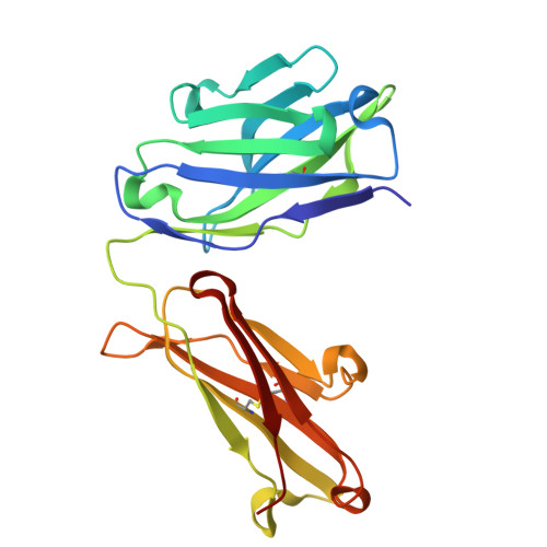
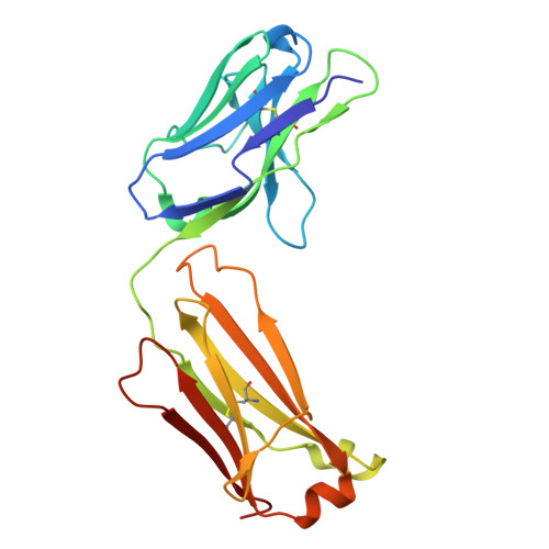
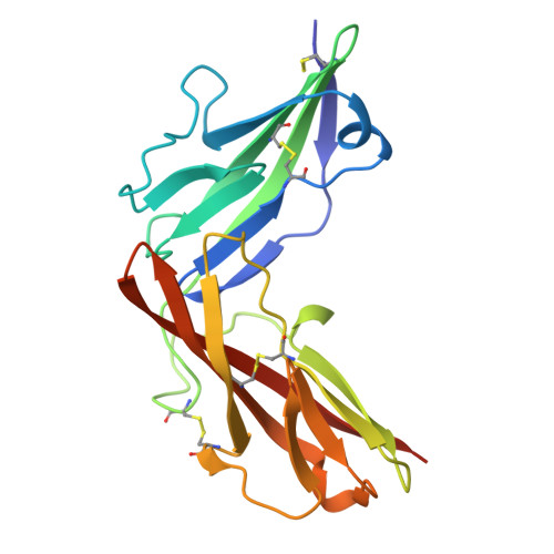
    Interleukin-36 receptor (IL-36R), belonging to the IL-1 receptor family, is crucial for host defense and tissue repair. Targeting cytokine receptors with low molecular weight (LMW) compounds remains challenging due to their interaction with the large surface area of cytokine. In this study, two encoded library technologies are used to identify LMW molecules binding to IL-36R's extracellular domain. The mRNA-based display technique identifies 36R-P138, a macrocyclic peptide blocking IL-36R signaling. Importantly, its optimized analog (36R-P192) also effectively suppresses expression of marker genes induced by IL-36 in human skin biopsies. DNA encoded libraries (DEL) screening delivers 36R-D481, a high affinity LMW IL-36R binder, effectively inhibiting IL-36 signaling. X-ray crystallography analysis reveals that both the cyclic peptide and DEL-compound bind to the IL-36R's D1 domain, potentially disrupting IL-36 cytokine binding. This study demonstrates that it is possible to target a cytokine receptor within the IL-1 receptor family using a small molecule ( < 1000 Da).


  • Organizational Affiliation
    • Novartis Biomedical Research, Novartis Campus, CH-4002, Basel, Switzerland. juraj.velcicky@novartis.com.

Macromolecules
Find similar proteins by:  (by identity cutoff)  |  3D Structure
Entity ID: 1
MoleculeChains Sequence LengthOrganismDetailsImage
BI00655130 Fab heavy chainA [auth H]221Homo sapiensMutation(s): 0 
Entity Groups  
Sequence Clusters30% Identity50% Identity70% Identity90% Identity95% Identity100% Identity
Sequence Annotations
Expand
  • Reference Sequence
Find similar proteins by:  (by identity cutoff)  |  3D Structure
Entity ID: 2
MoleculeChains Sequence LengthOrganismDetailsImage
BI00655130 Fab light chainB [auth L]215Homo sapiensMutation(s): 0 
Entity Groups  
Sequence Clusters30% Identity50% Identity70% Identity90% Identity95% Identity100% Identity
Sequence Annotations
Expand
  • Reference Sequence
Find similar proteins by:  (by identity cutoff)  |  3D Structure
Entity ID: 3
MoleculeChains Sequence LengthOrganismDetailsImage
Interleukin-1 receptor-like 2C [auth R]207Homo sapiensMutation(s): 0 
Gene Names: IL1RL2IL1RRP2
EC: 3.2.2.6
UniProt & NIH Common Fund Data Resources
Find proteins for Q9HB29 (Homo sapiens)
Explore Q9HB29 
Go to UniProtKB:  Q9HB29
PHAROS:  Q9HB29
GTEx:  ENSG00000115598 
Entity Groups  
Sequence Clusters30% Identity50% Identity70% Identity90% Identity95% Identity100% Identity
UniProt GroupQ9HB29
Glycosylation
Glycosylation Sites: 2Go to GlyGen: Q9HB29-1
Sequence Annotations
Expand
  • Reference Sequence
Oligosaccharides

Help

Entity ID: 4
MoleculeChains Length2D Diagram Glycosylation3D Interactions
2-acetamido-2-deoxy-beta-D-glucopyranose-(1-4)-2-acetamido-2-deoxy-beta-D-glucopyranoseD [auth A]2N-Glycosylation
Glycosylation Resources
GlyTouCan:  G42666HT
GlyCosmos:  G42666HT
GlyGen:  G42666HT
Small Molecules
Ligands 3 Unique
IDChains Name / Formula / InChI Key2D Diagram3D Interactions
A1H62 (Subject of Investigation/LOI)
Query on A1H62

Download Ideal Coordinates CCD File 
G [auth R]4-[3-[[(1~{S})-1-(3-bromophenyl)-3-[2-(2-morpholin-4-ylethoxy)ethylamino]-3-oxidanylidene-propyl]carbamoyl]-1,4,6,7-tetrahydropyrazolo[4,3-c]pyridin-5-yl]-5-cyano-thieno[2,3-b]pyridine-2-carboxylic acid
C33 H35 Br N8 O6 S
RJANKZNKWUTXJN-SANMLTNESA-N
NAG
Query on NAG

Download Ideal Coordinates CCD File 
F [auth R]2-acetamido-2-deoxy-beta-D-glucopyranose
C8 H15 N O6
OVRNDRQMDRJTHS-FMDGEEDCSA-N
PO4
Query on PO4

Download Ideal Coordinates CCD File 
E [auth H]PHOSPHATE ION
O4 P
NBIIXXVUZAFLBC-UHFFFAOYSA-K
Experimental Data & Validation

Experimental Data

  • Method: X-RAY DIFFRACTION
  • Resolution: 2.40 Å
  • R-Value Free:  0.216 (Depositor), 0.205 (DCC) 
  • R-Value Work:  0.189 (Depositor), 0.177 (DCC) 
  • R-Value Observed: 0.190 (Depositor) 
Space Group: P 21 21 21
Unit Cell:
Length ( Å )Angle ( ˚ )
a = 60.55α = 90
b = 69.94β = 90
c = 232.83γ = 90
Software Package:
Software NamePurpose
BUSTERrefinement
autoPROCdata reduction
STARANISOdata scaling
PHASERphasing

Structure Validation

View Full Validation Report


Ligand Structure Quality Assessment 


Entry History & Funding Information

Deposition Data


Funding OrganizationLocationGrant Number
Not funded--

Revision History  (Full details and data files)

  • Version 1.0: 2025-02-26
    Type: Initial release