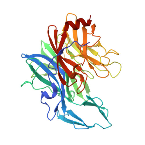
9EQQ | pdb_00009eqq

Uhgb_MS mannoside synthase from an unknown human gut bacterium in complex with Mannose


Experimental Data Snapshot

  • Method: X-RAY DIFFRACTION
  • Resolution: 2.55 Å
  • R-Value Free: 
    0.207 (Depositor), 0.208 (DCC) 
  • R-Value Work: 
    0.179 (Depositor), 0.187 (DCC) 

Starting Model: in silico
View more details

wwPDB Validation 3D Report Full Report

Validation slider image for 9EQQ

Ligand Structure Quality Assessment 


This is version 1.1 of the entry. See complete history. 

Literature

Dual-Activity Mannosyltransferase Phosphorylases in Family 130 of Glycoside Hydrolases.

Ladeveze, S., Tarquis, L., Cioci, G., Guieysse, D., Durand, J., Faure, R., Li, A., Esque, J., Vivier, M., Malbert, Y., Laville, E., Potocki-Veronese, G.

(2026) ACS Catal 16: 1211-1223

  • DOI: https://doi.org/10.1021/acscatal.5c06782
  • Primary Citation Related Structures: 
    9EQQ, 9FJ8, 9GPH

  • PubMed Abstract: 

    We discovered that certain mannoside-active enzymes from family GH130 possess both glycoside phosphorylase (GP) and weak glycosyltransferase (GT) activities. This dual activity was observed only in glycoside phosphorylases acting on β-(1,2)- and β-(1,3)-mannosides, being absent in β-(1,4)-mannoside phosphorylases. We provide several three-dimensional (3D) structures of β-(1,2)-mannosyltransferase phosphorylases Uhgb_MS (identified from the human gut microbiome (subfamily GH130_4)) and Teth514_1788 (from Thermoanaerobacter sp. X514 (subfamily GH130_6)) and analyzed GT/GP partitioning among enzymes acting on d-mannosides. Structural analysis and mutagenesis of Uhgb_MS allowed us to show that loop L2 folding, as well as nucleotide and phosphate binding residues (P206, N211, R242) at the location of the common structural phosphate of αMan1P and GDP-Man, is an important structural element of bifunctionality in GH130.


  • Organizational Affiliation: 
    • TBI, Université de Toulouse, CNRS, INRAE, INSA, 31077 Toulouse, France.

Macromolecule Content 

  • Total Structure Weight: 39.42 kDa 
  • Atom Count: 2,909 
  • Modeled Residue Count: 347 
  • Deposited Residue Count: 350 
  • Unique protein chains: 1

Macromolecules

Find similar proteins by:|  3D Structure
Entity ID: 1
MoleculeChains  Sequence LengthOrganismDetailsImage
Uhgb_MS mannoside synthase from an unknown human gut bacterium350metagenomeMutation(s): 0 
UniProt
Find proteins for F0QZL2 (Phocaeicola salanitronis (strain DSM 18170 / JCM 13657 / CCUG 60908 / BL78))
Explore F0QZL2 
Go to UniProtKB:  F0QZL2
Entity Groups
Sequence Clusters30% Identity50% Identity70% Identity90% Identity95% Identity100% Identity
UniProt GroupF0QZL2
Sequence Annotations
Expand
Reference Sequence

Experimental Data & Validation

Experimental Data

  • Method: X-RAY DIFFRACTION
  • Resolution: 2.55 Å
  • R-Value Free:  0.207 (Depositor), 0.208 (DCC) 
  • R-Value Work:  0.179 (Depositor), 0.187 (DCC) 
Space Group: P 61 2 2
Unit Cell:
Length ( Å )Angle ( ˚ )
a = 176.629α = 90
b = 176.629β = 90
c = 83.539γ = 120
Software Package:
Software NamePurpose
REFMACrefinement
XDSdata reduction
XDSdata scaling
MOLREPphasing

Structure Validation

View Full Validation Report


Ligand Structure Quality Assessment 


Entry History 

& Funding Information

Deposition Data


Funding OrganizationLocationGrant Number
Not funded--

Revision History  (Full details and data files)

  • Version 1.0: 2025-10-08
    Type: Initial release
  • Version 1.1: 2026-09-30
    Changes: Database references