9EKF | pdb_00009ekf

CryoEM structure of H5N1 A/Texas/37/2024 HA bound to Fab 65C6 and an auto glycan occupying the receptor-binding site


Experimental Data Snapshot

  • Method: ELECTRON MICROSCOPY
  • Resolution: 2.68 Å
  • Aggregation State: PARTICLE 
  • Reconstruction Method: SINGLE PARTICLE 

wwPDB Validation 3D Report Full Report

Validation slider image for 9EKF

This is version 1.2 of the entry. See complete history

Literature

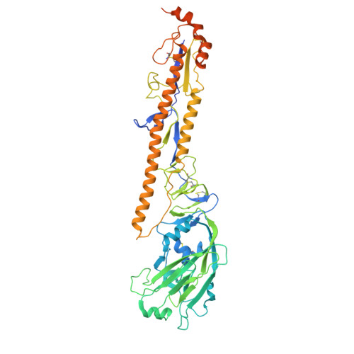
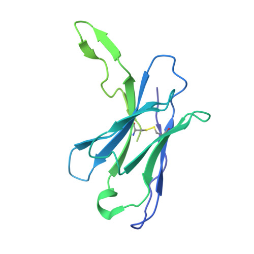
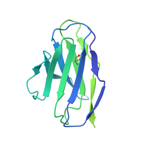
Structure of a zoonotic H5N1 hemagglutinin reveals a receptor-binding site occupied by an auto-glycan.

Morano, N.C.Guo, Y.Becker, J.E.Li, Z.Yu, J.Ho, D.D.Shapiro, L.Kwong, P.D.

(2025) Structure 33: 228-233.e3

  • DOI: https://doi.org/10.1016/j.str.2025.01.001
  • Primary Citation Related Structures: 
    9EKF

  • PubMed Abstract: 

    Highly pathogenic avian influenza has spilled into many mammals, most notably cows and poultry, with several dozen human breakthrough infections. Zoonotic crossovers, with hemagglutinins mutated to enhance viral ability to use human α2-6-linked sialic acid receptors versus avian α2-3-linked ones, highlight the pandemic risk. To gain insight into these crossovers, we determined the cryoelectron microscopy (cryo-EM) structure of the hemagglutinin from the zoonotic H5N1 A/Texas/37/2024 strain (clade 2.3.4.4b) in complex with a previously reported neutralizing antibody. Surprisingly, we found that the receptor-binding site of this H5N1 hemagglutinin was already occupied by an α2-3-linked sialic acid and that this glycan emanated from asparagine N169 of a neighboring protomer on hemagglutinin itself. This structure thus highlights recognition by influenza hemagglutinin of an "auto"-α2-3-linked sialic acid from N169, an N-linked glycan conserved in 95% of H5 strains, and adds "auto-glycan recognition," which may play a role in viral dispersal, to the complexities surrounding H5N1 zoonosis.


  • Organizational Affiliation
    • Aaron Diamond AIDS Research Center, Columbia University Vagelos College of Physicians and Surgeons, New York, NY 10032, USA; Zuckerman Mind Brain Behavior Institute, Columbia University, New York, NY 10027, USA; Department of Biochemistry and Molecular Biophysics, Columbia University, New York, NY 10027, USA.

Macromolecule Content 

  • Total Structure Weight: 351.01 kDa 
  • Atom Count: 17,537 
  • Modeled Residue Count: 2,145 
  • Deposited Residue Count: 3,081 
  • Unique protein chains: 3

Macromolecules

Find similar proteins by:|  3D Structure
Entity ID: 1
MoleculeChains  Sequence LengthOrganismDetailsImage
Hemagglutinin
A, B, C
576Influenza A virusMutation(s): 0 
UniProt
Find proteins for A0A8E4ZAK5 (Influenza A virus)
Explore A0A8E4ZAK5 
Go to UniProtKB:  A0A8E4ZAK5
Entity Groups
Sequence Clusters30% Identity50% Identity70% Identity90% Identity95% Identity100% Identity
UniProt GroupA0A8E4ZAK5
Glycosylation
Glycosylation Sites: 4
Sequence Annotations
Expand
Reference Sequence
Find similar proteins by:|  3D Structure
Entity ID: 2
MoleculeChains  Sequence LengthOrganismDetailsImage
Fab 65C6 Heavy ChainD,
E [auth G],
F [auth H]
237Homo sapiensMutation(s): 0 
Find similar proteins by:|  3D Structure
Entity ID: 3
MoleculeChains  Sequence LengthOrganismDetailsImage
Fab 65C6 Light ChainG [auth I],
H [auth K],
I [auth L]
214Homo sapiensMutation(s): 0 

Oligosaccharides

Help  
Entity ID: 4
MoleculeChains Length2D Diagram GlycosylationD Interactions
N-acetyl-alpha-neuraminic acid-(2-3)-beta-D-galactopyranose-(1-4)-2-acetamido-2-deoxy-beta-D-glucopyranose-(1-2)-alpha-D-mannopyranose-(1-3)-beta-D-mannopyranose-(1-4)-2-acetamido-2-deoxy-beta-D-glucopyranose-(1-4)-2-acetamido-2-deoxy-beta-D-glucopyranoseJ [auth E],
K [auth F],
L [auth J]
7N/A
Glycosylation Resources
GlyTouCan: G85526ZL
GlyCosmos: G85526ZL
GlyGen: G85526ZL

Small Molecules

Ligands 1 Unique
IDChains Name / Formula / InChI Key2D Diagram3D Interactions
NAG

Query on NAG


Download:Ideal Coordinates CCD File
M [auth A]
N [auth A]
O [auth A]
P [auth A]
Q [auth B]
M [auth A],
N [auth A],
O [auth A],
P [auth A],
Q [auth B],
R [auth B],
S [auth B],
T [auth B],
U [auth C],
V [auth C],
W [auth C],
X [auth C]
2-acetamido-2-deoxy-beta-D-glucopyranose
C8 H15 N O6
OVRNDRQMDRJTHS-FMDGEEDCSA-N

Experimental Data & Validation

Experimental Data

  • Method: ELECTRON MICROSCOPY
  • Resolution: 2.68 Å
  • Aggregation State: PARTICLE 
  • Reconstruction Method: SINGLE PARTICLE 
EM Software:
TaskSoftware PackageVersion
RECONSTRUCTIONcryoSPARC

Structure Validation

View Full Validation Report


Entry History 

& Funding Information

Deposition Data


Funding OrganizationLocationGrant Number
National Institutes of Health/National Institute Of Allergy and Infectious Diseases (NIH/NIAID)United States--

Revision History  (Full details and data files)

  • Version 1.0: 2024-12-18
    Type: Initial release
  • Version 1.1: 2025-02-12
    Changes: Data collection, Database references
  • Version 1.2: 2025-02-19
    Changes: Data collection, Database references