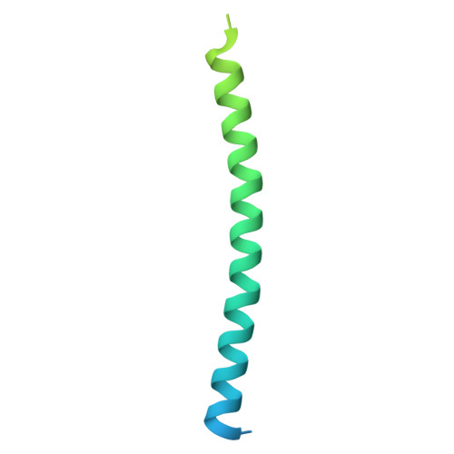
Nanobodies against Cavin1 reveal structural flexibility and regulated interactions of its N-terminal coiled-coil domain.
Gao, Y., Tillu, V.A., Wu, Y., Rae, J., Hall, T.E., Chen, K.E., Weeratunga, S., Guo, Q., Livingstone, E., Tham, W.H., Parton, R.G., Collins, B.M.(2025) J Cell Sci 138
- PubMed: 40260863 Search on PubMedSearch on PubMed Central
- DOI: https://doi.org/10.1242/jcs.263756
- Primary Citation Related Structures:
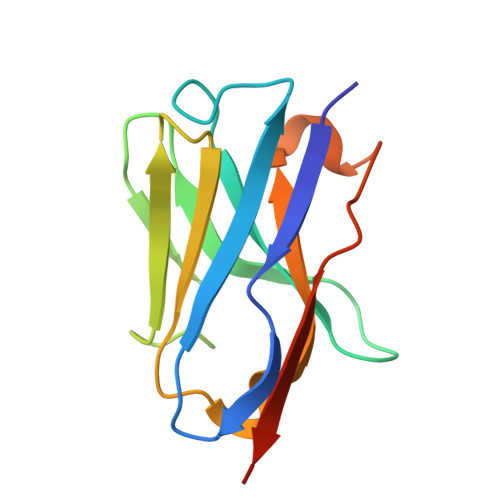
9EG6, 9EGN, 9EIU - PubMed Abstract:
Caveolae are abundant plasma membrane structures that regulate signalling, membrane homeostasis and mechanoprotection. Their formation is driven by caveolins and cavins and their coordinated interactions with lipids. Here, we developed nanobodies against the trimeric HR1 coiled-coil domain of Cavin1. We identified specific nanobodies that do not perturb Cavin1 membrane binding and localise to caveolae when expressed in cells. The crystal structure of a nanobody-Cavin 1 HR1 complex reveals a symmetric 3:3 architecture as validated by mutagenesis. In this structure, the C-terminal half of the HR1 domain is disordered, suggesting that the nanobody stabilises an open conformation of Cavin1, which has previously been identified as important for membrane interactions. A phosphomimic mutation in a threonine-serine pair proximal to this region reveals selective regulation of Cavin2 and Cavin3 association. These studies provide new insights into cavin domains required for assembly of multiprotein caveolar assemblies and describe new nanobody tools for structural and functional studies of caveolae.
- Institute for Molecular Bioscience, The University of Queensland, St. Lucia, Queensland 4067, Australia.
Organizational Affiliation: