9ECE | pdb_00009ece

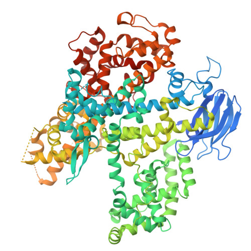
Inactive state of wild-type EsCas13d ternary complex with C4A mismatch


Experimental Data Snapshot

  • Method: ELECTRON MICROSCOPY
  • Resolution: 3.01 Å
  • Aggregation State: PARTICLE 
  • Reconstruction Method: SINGLE PARTICLE 

wwPDB Validation 3D Report Full Report

Validation slider image for 9ECE

This is version 1.1 of the entry. See complete history

Literature

Structural basis for target discrimination and activation by Cas13d.

Chou, C.W.Sinan, S.Kuo, H.C.Chang, Y.C.Arguello, C.Sahaya, D.Russell, R.Finkelstein, I.J.

(2026) Sci Adv 12: eaec4221-eaec4221

  • DOI: https://doi.org/10.1126/sciadv.aec4221
  • Primary Citation Related Structures: 
    9EBU, 9EC9, 9ECA, 9ECB, 9ECC, 9ECD, 9ECE

  • PubMed Abstract: 

    CRISPR-Cas13d is increasingly used for RNA knockdowns, but off-target cleavage of near-cognate RNAs hinders its broader adoption. Here, we solve seven cryo-electron microscopy structures of wild-type Cas13d in complex with matched and mismatched targets. These structures reveal active, intermediate, and inactive states that illustrate a detailed activation mechanism. Upon target RNA binding, the CRISPR RNA undergoes marked conformational changes. The Helical-1 domain transitions from a docked state with the amino-terminal domain to an allosterically switched conformation that stabilizes the RNA duplex. Quantitative kinetics show that a single proximal mismatch preserves the binding rate constant but abolishes nuclease activity by trapping Cas13d in an inactive state. We also identify an active site loop in the higher eukaryotes and prokaryotes nucleotide-binding (HEPN) domains that regulates substrate accessibility and can be mutated to generate both hypo- and hyperactivated variants. These findings establish the structural basis for Cas13d mismatch surveillance and provide a framework for engineering HEPN nuclease specificity and activity.


  • Organizational Affiliation
    • Department of Molecular Biosciences, The University of Texas at Austin, Austin, TX 78712, USA.

Macromolecule Content 

  • Total Structure Weight: 137.21 kDa 
  • Atom Count: 8,298 
  • Modeled Residue Count: 903 
  • Deposited Residue Count: 1,036 
  • Unique protein chains: 1
  • Unique nucleic acid chains: 2

Macromolecules


Find similar proteins by:|  3D Structure
Entity ID: 1
MoleculeChains  Sequence LengthOrganismDetailsImage
EsCas13d954[Eubacterium] siraeum DSM 15702Mutation(s): 0 
Gene Names: EUBSIR_02687
UniProt
Find proteins for B0MS50 ([Eubacterium] siraeum DSM 15702)
Explore B0MS50 
Go to UniProtKB:  B0MS50
Entity Groups
Sequence Clusters30% Identity50% Identity70% Identity90% Identity95% Identity100% Identity
UniProt GroupB0MS50
Sequence Annotations
Expand
Reference Sequence
Find similar nucleic acids by:  (by identity cutoff) 
Entity ID: 2
MoleculeChains LengthOrganismImage
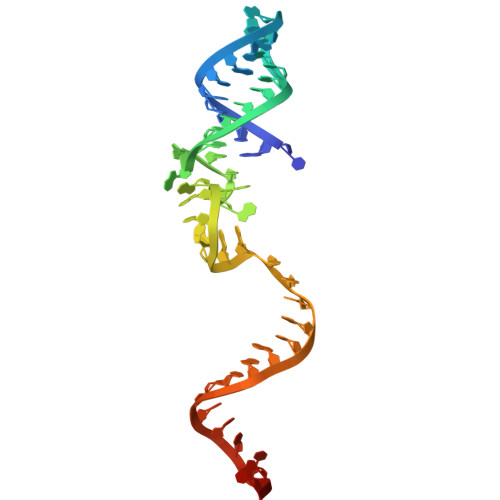
crRNA52[Eubacterium] siraeum DSM 15702
Sequence Annotations
Expand
Reference Sequence
Find similar nucleic acids by:  (by identity cutoff) 
Entity ID: 3
MoleculeChains LengthOrganismImage
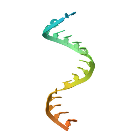
Target RNA with C4A mismatch30[Eubacterium] siraeum DSM 15702
Sequence Annotations
Expand
Reference Sequence

Small Molecules

Ligands 1 Unique
IDChains Name / Formula / InChI Key2D Diagram3D Interactions
MG

Query on MG


Download:Ideal Coordinates CCD File
D [auth B]MAGNESIUM ION
Mg
JLVVSXFLKOJNIY-UHFFFAOYSA-N

Experimental Data & Validation

Experimental Data

  • Method: ELECTRON MICROSCOPY
  • Resolution: 3.01 Å
  • Aggregation State: PARTICLE 
  • Reconstruction Method: SINGLE PARTICLE 
EM Software:
TaskSoftware PackageVersion
MODEL REFINEMENTPHENIX1.21.2_5419

Structure Validation

View Full Validation Report


Entry History 

& Funding Information

Deposition Data


Funding OrganizationLocationGrant Number
Welch FoundationUnited StatesF-1808

Revision History  (Full details and data files)

  • Version 1.0: 2025-11-19
    Type: Initial release
  • Version 1.1: 2026-09-02
    Changes: Data collection, Database references