9EBI | pdb_00009ebi

Human adenosine A3 receptor Gi complex (mini-Gsi chimera) bound to Piclidenoson (CF101, IB-MECA)


Experimental Data Snapshot

  • Method: ELECTRON MICROSCOPY
  • Resolution: 3.60 Å
  • Aggregation State: PARTICLE 
  • Reconstruction Method: SINGLE PARTICLE 

wwPDB Validation   3D Report Full Report


This is version 1.1 of the entry. See complete history


Literature

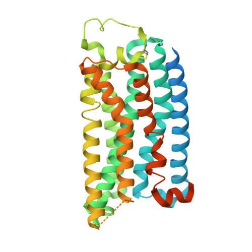
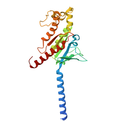
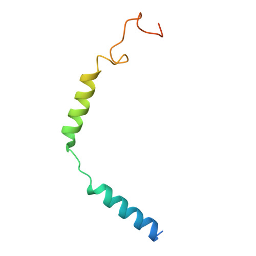
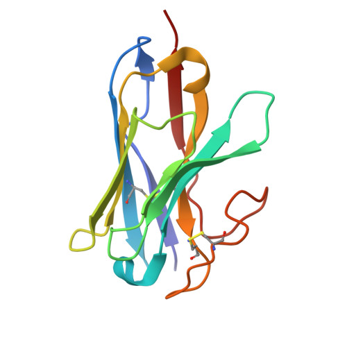
Molecular basis of ligand binding and receptor activation at the human A 3 adenosine receptor.

Zhang, L.Mobbs, J.I.Bennetts, F.M.Venugopal, H.Nguyen, A.T.N.Christopoulos, A.van der Es, D.Heitman, L.H.May, L.T.Glukhova, A.Thal, D.M.

(2025) Nat Commun 16: 7674-7674

  • DOI: https://doi.org/10.1038/s41467-025-62872-x
  • Primary Citation of Related Structures:  
    9EBH, 9EBI, 9EHS

  • PubMed Abstract: 

    Adenosine receptors (ARs: A 1 AR, A 2A AR, A 2B AR, and A 3 AR) are crucial therapeutic targets; however, developing selective, efficacious drugs for them remains a significant challenge. Here, we present high-resolution cryo-electron microscopy (cryo-EM) structures of the human A 3 AR in three distinct functional states: bound to the endogenous agonist adenosine, the clinically relevant agonist Piclidenoson, and the covalent antagonist LUF7602. These structures, complemented by mutagenesis and pharmacological studies, reveal an A 3 AR activation mechanism that involves an extensive hydrogen bond network from the extracellular surface down to the orthosteric binding site. In addition, we identify a cryptic pocket that accommodates the N 6 -iodobenzyl group of Piclidenoson through a ligand-dependent conformational change of M174 5.35 . Our comprehensive structural and functional characterisation of A 3 AR advances our understanding of adenosine receptor pharmacology and establishes a foundation for developing more selective therapeutics for various disorders, including inflammatory diseases, cancer, and glaucoma.


  • Organizational Affiliation
    • Drug Discovery Biology, Monash Institute of Pharmaceutical Sciences, Monash University, Parkville, VIC, Australia.

Macromolecules
Find similar proteins by:  (by identity cutoff)  |  3D Structure
Entity ID: 1
MoleculeChains Sequence LengthOrganismDetailsImage
Adenosine receptor A3A [auth R]363Homo sapiensMutation(s): 0 
Gene Names: ADORA3
UniProt & NIH Common Fund Data Resources
Find proteins for P0DMS8 (Homo sapiens)
Explore P0DMS8 
Go to UniProtKB:  P0DMS8
PHAROS:  P0DMS8
GTEx:  ENSG00000282608 
Entity Groups  
Sequence Clusters30% Identity50% Identity70% Identity90% Identity95% Identity100% Identity
UniProt GroupP0DMS8
Sequence Annotations
Expand
  • Reference Sequence
Find similar proteins by:  (by identity cutoff)  |  3D Structure
Entity ID: 2
MoleculeChains Sequence LengthOrganismDetailsImage
mini-Gs/i chimeraB [auth A]257Homo sapiensMutation(s): 0 
Entity Groups  
Sequence Clusters30% Identity50% Identity70% Identity90% Identity95% Identity100% Identity
Sequence Annotations
Expand
  • Reference Sequence
Find similar proteins by:  (by identity cutoff)  |  3D Structure
Entity ID: 3
MoleculeChains Sequence LengthOrganismDetailsImage
Guanine nucleotide-binding protein G(I)/G(S)/G(T) subunit beta-1C [auth B]349Homo sapiensMutation(s): 0 
Gene Names: GNB1
UniProt & NIH Common Fund Data Resources
Find proteins for P62873 (Homo sapiens)
Explore P62873 
Go to UniProtKB:  P62873
PHAROS:  P62873
GTEx:  ENSG00000078369 
Entity Groups  
Sequence Clusters30% Identity50% Identity70% Identity90% Identity95% Identity100% Identity
UniProt GroupP62873
Sequence Annotations
Expand
  • Reference Sequence
Find similar proteins by:  (by identity cutoff)  |  3D Structure
Entity ID: 4
MoleculeChains Sequence LengthOrganismDetailsImage
Guanine nucleotide-binding protein G(I)/G(S)/G(O) subunit gamma-2D [auth G]70Homo sapiensMutation(s): 0 
Gene Names: GNG2
UniProt & NIH Common Fund Data Resources
Find proteins for P59768 (Homo sapiens)
Explore P59768 
Go to UniProtKB:  P59768
PHAROS:  P59768
GTEx:  ENSG00000186469 
Entity Groups  
Sequence Clusters30% Identity50% Identity70% Identity90% Identity95% Identity100% Identity
UniProt GroupP59768
Sequence Annotations
Expand
  • Reference Sequence
Find similar proteins by:  (by identity cutoff)  |  3D Structure
Entity ID: 5
MoleculeChains Sequence LengthOrganismDetailsImage
Nb35E [auth N]128Lama glamaMutation(s): 0 
Entity Groups  
Sequence Clusters30% Identity50% Identity70% Identity90% Identity95% Identity100% Identity
Sequence Annotations
Expand
  • Reference Sequence
Small Molecules
Ligands 1 Unique
IDChains Name / Formula / InChI Key2D Diagram3D Interactions
Q8L (Subject of Investigation/LOI)
Query on Q8L

Download Ideal Coordinates CCD File 
F [auth R]Piclidenoson
C18 H19 I N6 O4
HUJXGQILHAUCCV-MOROJQBDSA-N
Experimental Data & Validation

Experimental Data

  • Method: ELECTRON MICROSCOPY
  • Resolution: 3.60 Å
  • Aggregation State: PARTICLE 
  • Reconstruction Method: SINGLE PARTICLE 
EM Software:
TaskSoftware PackageVersion
MODEL REFINEMENTPHENIX1.21rc1_5127

Structure Validation

View Full Validation Report


Entry History & Funding Information

Deposition Data


Funding OrganizationLocationGrant Number
National Health and Medical Research Council (NHMRC, Australia)Australia1196951
National Health and Medical Research Council (NHMRC, Australia)Australia138448
Australian Research Council (ARC)AustraliaLP180100560

Revision History  (Full details and data files)

  • Version 1.0: 2025-08-20
    Type: Initial release
  • Version 1.1: 2025-09-03
    Changes: Data collection, Database references