9E8P | pdb_00009e8p

Crystal Structure of GABARAP-ATG3 conjugate

  • Classification: LIGASE
  • Organism(s): Homo sapiens
  • Expression System: Escherichia coli
  • Mutation(s): Yes 

  • Deposited: 2024-11-05 Released: 2025-08-06 
  • Deposition Author(s): Ohashi, K., Otomo, T.
  • Funding Organization(s): National Institutes of Health/National Institute of General Medical Sciences (NIH/NIGMS)

Experimental Data Snapshot

  • Method: X-RAY DIFFRACTION
  • Resolution: 2.70 Å
  • R-Value Free: 
    0.249 (Depositor), 0.247 (DCC) 
  • R-Value Work: 
    0.218 (Depositor), 0.218 (DCC) 
  • R-Value Observed: 
    0.221 (Depositor) 

Starting Models: experimental
View more details

wwPDB Validation   3D Report Full Report


This is version 1.1 of the entry. See complete history


Literature

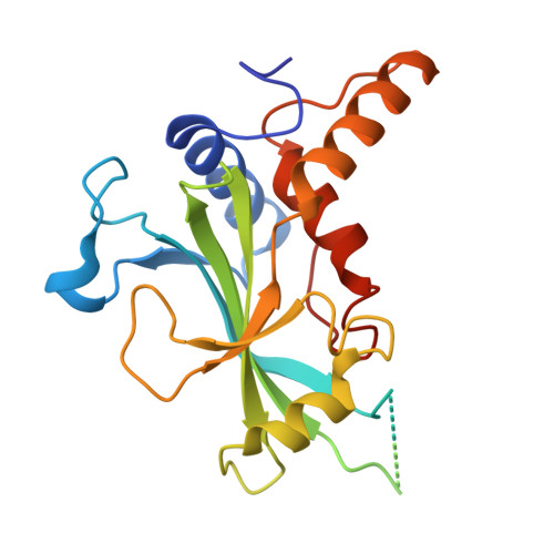
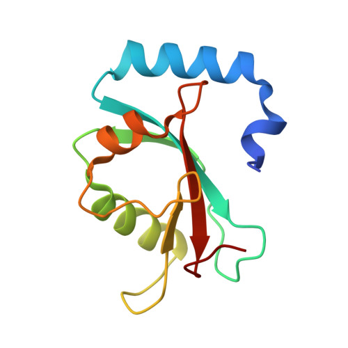
Structural Insights into the GABARAP-ATG3 Backside Interaction and Apo ATG3 Conformation.

Ohashi, K.Kroon, G.J.Otomo, T.

(2025) Biochemistry 64: 3178-3189

  • DOI: https://doi.org/10.1021/acs.biochem.4c00485
  • Primary Citation of Related Structures:  
    9E8P

  • PubMed Abstract: 

    Members of the ATG8 family of ubiquitin-like proteins (Ubls) are covalently attached to phosphatidylethanolamine (PE) on nascent autophagosomal membranes, where they recruit cargo receptors and promote membrane expansion. Although the overall lipidation pathway is well established, the molecular details-particularly those involving the E2 enzyme ATG3-remain incompletely defined. Here, we uncover a previously unrecognized, noncovalent binding mode between the mammalian ATG8 protein GABARAP and the backside of ATG3's catalytic E2 domain. In crystals, an isopeptide-linked GABARAP∼ATG3 conjugate self-assembles into a helical filament via this backside interface, mirroring architectures observed for canonical Ub/Ubl∼E2 conjugates. The E2 backside-binding surface on GABARAP is topologically distinct from those of other Ub/Ubl proteins and overlaps the LC3-interacting region (LIR) motif-binding site. Solution NMR confirms this interaction, and targeted mutagenesis shows that disrupting the interface impairs PE conjugation. Complementary NMR and AlphaFold modeling of apo ATG3 reveal an intramolecular contact between a segment of its flexible region (FR) and the catalytic core that suppresses conjugation. Together, these findings establish backside engagement as a critical feature of ATG8 lipidation and illuminate the dynamic architecture and regulation of ATG3.


  • Organizational Affiliation
    • Department of Integrative Structural and Computational Biology, The Scripps Research Institute, 10550 North Torrey Pines Rd, La Jolla, California 92037, United States.

Macromolecules
Find similar proteins by:  (by identity cutoff)  |  3D Structure
Entity ID: 1
MoleculeChains Sequence LengthOrganismDetailsImage
Ubiquitin-like-conjugating enzyme ATG3
A, B
223Homo sapiensMutation(s): 1 
Gene Names: ATG3APG3APG3L
EC: 2.3.2
UniProt & NIH Common Fund Data Resources
Find proteins for Q9NT62 (Homo sapiens)
Explore Q9NT62 
Go to UniProtKB:  Q9NT62
PHAROS:  Q9NT62
GTEx:  ENSG00000144848 
Entity Groups  
Sequence Clusters30% Identity50% Identity70% Identity90% Identity95% Identity100% Identity
UniProt GroupQ9NT62
Sequence Annotations
Expand
  • Reference Sequence
Find similar proteins by:  (by identity cutoff)  |  3D Structure
Entity ID: 2
MoleculeChains Sequence LengthOrganismDetailsImage
Gamma-aminobutyric acid receptor-associated protein
C, D
118Homo sapiensMutation(s): 0 
Gene Names: GABARAPFLC3BHT004
UniProt & NIH Common Fund Data Resources
Find proteins for O95166 (Homo sapiens)
Explore O95166 
Go to UniProtKB:  O95166
PHAROS:  O95166
GTEx:  ENSG00000170296 
Entity Groups  
Sequence Clusters30% Identity50% Identity70% Identity90% Identity95% Identity100% Identity
UniProt GroupO95166
Sequence Annotations
Expand
  • Reference Sequence
Experimental Data & Validation

Experimental Data

  • Method: X-RAY DIFFRACTION
  • Resolution: 2.70 Å
  • R-Value Free:  0.249 (Depositor), 0.247 (DCC) 
  • R-Value Work:  0.218 (Depositor), 0.218 (DCC) 
  • R-Value Observed: 0.221 (Depositor) 
Space Group: P 43 21 2
Unit Cell:
Length ( Å )Angle ( ˚ )
a = 97.388α = 90
b = 97.388β = 90
c = 166.774γ = 90
Software Package:
Software NamePurpose
PHENIXrefinement
XDSdata reduction
XSCALEdata scaling
PHENIXphasing

Structure Validation

View Full Validation Report


Entry History & Funding Information

Deposition Data


Funding OrganizationLocationGrant Number
National Institutes of Health/National Institute of General Medical Sciences (NIH/NIGMS)United StatesR01GM092740

Revision History  (Full details and data files)

  • Version 1.0: 2025-08-06
    Type: Initial release
  • Version 1.1: 2025-08-20
    Changes: Database references