9E8A | pdb_00009e8a

Integrin aIIbb3 bent conformation from human platelet membrane crude preparation

  • Classification: BLOOD CLOTTING
  • Organism(s): Homo sapiens
  • Mutation(s): No 

  • Deposited: 2024-11-05 Released: 2025-10-15 
  • Deposition Author(s): Han, X., Nieman, M.T.
  • Funding Organization(s): National Institutes of Health/National Heart, Lung, and Blood Institute (NIH/NHLBI)

Experimental Data Snapshot

  • Method: ELECTRON MICROSCOPY
  • Resolution: 2.75 Å
  • Aggregation State: PARTICLE 
  • Reconstruction Method: SINGLE PARTICLE 

wwPDB Validation   3D Report Full Report


This is version 1.0 of the entry. See complete history


Literature

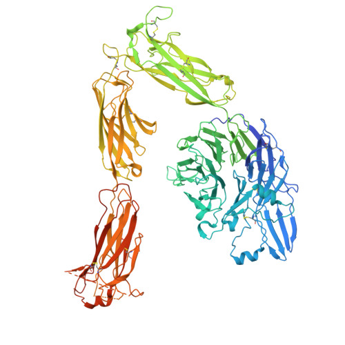
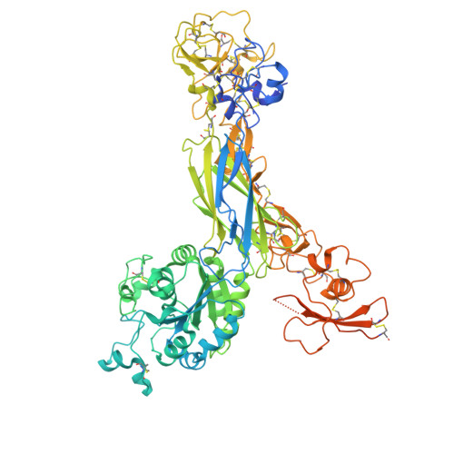
Elucidating the dynamics of integrin alpha IIb beta 3 from native platelet membranes by cryo-EM with build-and-retrieve method.

Han, X.Zhang, Z.Su, C.C.Lyu, M.Miyagi, M.Yu, E.Nieman, M.T.

(2025) Blood Adv 9: 4592-4606

  • DOI: https://doi.org/10.1182/bloodadvances.2025016209
  • Primary Citation of Related Structures:  
    9E8A, 9E8B, 9E8C

  • PubMed Abstract: 

    Platelets fulfill their essential physiological roles sensing the extracellular environment through their membrane proteins. The native membrane environment provides essential regulatory cues that affect the protein structure and mechanism of action. Single-particle cryogenic electron microscopy (cryo-EM) has transformed structural biology by allowing high-resolution structures of membrane proteins to be solved from homogeneous samples. Our recent breakthroughs in data processing now make it feasible to obtain atomic-level-resolution protein structures from crude preparations in their native environments by integrating cryo-EM with the "build-and-retrieve" (BaR) data processing methodology. We applied this iterative bottom-up methodology on resting human platelet membranes for an in-depth systems biology approach to uncover how lipids, metal binding, post-translational modifications, and cofactor associations in the native environment regulate platelet function at the molecular level. Here, we report using cryo-EM followed by the BaR method to solve the unmodified integrin αIIbβ3 structure directly from resting human platelet membranes in its inactivated and intermediate states at 2.75 and 2.67 Å, respectively. Furthermore, we also solved a novel dimer conformation of αIIbβ3 at 2.85 Å formed by 2 intermediate states of αIIbβ3. This may indicate a previously unknown self-regulatory mechanism of αIIbβ3 in its native environment. In conclusion, our data show the power of using cryo-EM with the BaR method to determine 3 distinct structures including a novel dimer directly from natural sources. This approach allows us to identify unrecognized regulation mechanisms for proteins without artifacts owing to purification processes. These data have the potential to enrich our understanding of platelet signaling circuitry.


  • Organizational Affiliation
    • Department of Pharmacology, School of Medicine, Case Western Reserve University, Cleveland, OH.

Macromolecules
Find similar proteins by:  (by identity cutoff)  |  3D Structure
Entity ID: 1
MoleculeChains Sequence LengthOrganismDetailsImage
Integrin alpha-IIb1,039Homo sapiensMutation(s): 0 
UniProt & NIH Common Fund Data Resources
Find proteins for P08514 (Homo sapiens)
Explore P08514 
Go to UniProtKB:  P08514
PHAROS:  P08514
GTEx:  ENSG00000005961 
Entity Groups  
Sequence Clusters30% Identity50% Identity70% Identity90% Identity95% Identity100% Identity
UniProt GroupP08514
Glycosylation
Glycosylation Sites: 3Go to GlyGen: P08514-1
Sequence Annotations
Expand
  • Reference Sequence
Find similar proteins by:  (by identity cutoff)  |  3D Structure
Entity ID: 2
MoleculeChains Sequence LengthOrganismDetailsImage
Integrin beta-3788Homo sapiensMutation(s): 0 
UniProt & NIH Common Fund Data Resources
Find proteins for P05106 (Homo sapiens)
Explore P05106 
Go to UniProtKB:  P05106
PHAROS:  P05106
GTEx:  ENSG00000259207 
Entity Groups  
Sequence Clusters30% Identity50% Identity70% Identity90% Identity95% Identity100% Identity
UniProt GroupP05106
Glycosylation
Glycosylation Sites: 5Go to GlyGen: P05106-1
Sequence Annotations
Expand
  • Reference Sequence

Find similar proteins by:  Sequence   |   3D Structure  

Entity ID: 3
MoleculeChains Sequence LengthOrganismDetailsImage
GLY-ARG-GLY-ASP4Homo sapiensMutation(s): 0 
Sequence Annotations
Expand
  • Reference Sequence
Oligosaccharides

Help

Entity ID: 4
MoleculeChains Length2D Diagram Glycosylation3D Interactions
2-acetamido-2-deoxy-beta-D-glucopyranose-(1-4)-2-acetamido-2-deoxy-beta-D-glucopyranose
D, E, F, G
2N-Glycosylation
Glycosylation Resources
GlyTouCan:  G42666HT
GlyCosmos:  G42666HT
GlyGen:  G42666HT
Entity ID: 5
MoleculeChains Length2D Diagram Glycosylation3D Interactions
beta-D-mannopyranose-(1-4)-2-acetamido-2-deoxy-beta-D-glucopyranose-(1-4)-2-acetamido-2-deoxy-beta-D-glucopyranose
H, I
3N-Glycosylation
Glycosylation Resources
GlyTouCan:  G15407YE
GlyCosmos:  G15407YE
GlyGen:  G15407YE
Entity ID: 6
MoleculeChains Length2D Diagram Glycosylation3D Interactions
2-acetamido-2-deoxy-beta-D-glucopyranose-(1-4)-2-acetamido-2-deoxy-beta-D-glucopyranose-(1-4)-beta-D-mannopyranose-(1-6)-beta-D-mannopyranose
J
4N/A
Small Molecules
Ligands 3 Unique
IDChains Name / Formula / InChI Key2D Diagram3D Interactions
NAG
Query on NAG

Download Ideal Coordinates CCD File 
O [auth A],
P [auth A],
R [auth A],
V [auth B]
2-acetamido-2-deoxy-beta-D-glucopyranose
C8 H15 N O6
OVRNDRQMDRJTHS-FMDGEEDCSA-N
CA
Query on CA

Download Ideal Coordinates CCD File 
K [auth A]
L [auth A]
M [auth A]
N [auth A]
Q [auth A]
K [auth A],
L [auth A],
M [auth A],
N [auth A],
Q [auth A],
T [auth B],
U [auth B]
CALCIUM ION
Ca
BHPQYMZQTOCNFJ-UHFFFAOYSA-N
MG
Query on MG

Download Ideal Coordinates CCD File 
S [auth B]MAGNESIUM ION
Mg
JLVVSXFLKOJNIY-UHFFFAOYSA-N
Experimental Data & Validation

Experimental Data

  • Method: ELECTRON MICROSCOPY
  • Resolution: 2.75 Å
  • Aggregation State: PARTICLE 
  • Reconstruction Method: SINGLE PARTICLE 
EM Software:
TaskSoftware PackageVersion
MODEL REFINEMENTPHENIX

Structure Validation

View Full Validation Report


Entry History & Funding Information

Deposition Data


Funding OrganizationLocationGrant Number
National Institutes of Health/National Heart, Lung, and Blood Institute (NIH/NHLBI)United StatesHL098217

Revision History  (Full details and data files)

  • Version 1.0: 2025-10-15
    Type: Initial release