9E6X | pdb_00009e6x

Crystal structure of ferritin


Experimental Data Snapshot

  • Method: X-RAY DIFFRACTION
  • Resolution: 2.50 Å
  • R-Value Free: 
    0.256 (Depositor), 0.256 (DCC) 
  • R-Value Work: 
    0.196 (Depositor), 0.196 (DCC) 
  • R-Value Observed: 
    0.202 (Depositor) 

Starting Model: experimental
View more details

wwPDB Validation 3D Report Full Report

Validation slider image for 9E6X

Ligand Structure Quality Assessment 


This is version 1.1 of the entry. See complete history

Literature

Processing of Freestanding Single Supercrystal Assembled by Atomically Precise Protein-Decorated Nanoparticles.

Huang, X.Huang, Q.Feng, S.Wang, Z.

(2025) Nano Lett 25: 7178-7185

  • DOI: https://doi.org/10.1021/acs.nanolett.5c01619
  • Primary Citation Related Structures: 
    9E6X

  • PubMed Abstract: 

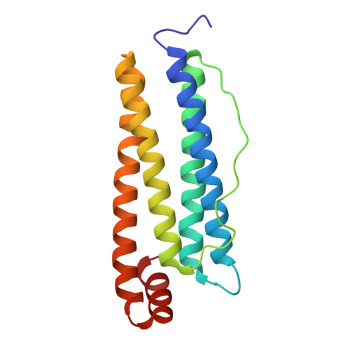
    Protein-decorated ferrihydrite nanoparticles (NPs) can self-assemble into periodically ordered structures with response to an external stimulus. X-ray crystallography determines the self-assembly of ferritin-folded nanocages into a face-centered cubic ( fcc ) structure at atomic resolution, while Cryo-EM imaging with 3D Ab-initio reconstruction defines the size, shape, and distribution of inside NPs at a subnanoscale. Processing of freestanding crystals reveals a large 3D spacing variation of 314% with preservation of translational symmetry. At an internanocage separation of 3.3 nm, crystals still maintain both translational and orientational ordering. In situ SAXS measurements reveal that the diffusion-driven NP assembly starts with a close separation to 3.3 nm. Compression of the single crystal leads to a 3D spacing reduction of ∼40%, giving a rigid modulus of 0.81 GPa. Enhanced stress causes water release from the ordered lattice and triggers biomolecule unfolding. This study provides insights for designing molecules to enhance directional interactions in the NP assembly and engineering control of active materials for adaptive applications.


  • Organizational Affiliation
    • Cornell High Energy Synchrotron Sources, Cornell University, Ithaca, New York 14853, United States.

Macromolecule Content 

  • Total Structure Weight: 20.23 kDa 
  • Atom Count: 1,397 
  • Modeled Residue Count: 170 
  • Deposited Residue Count: 170 
  • Unique protein chains: 1

Macromolecules

Find similar proteins by:|  3D Structure
Entity ID: 1
MoleculeChains  Sequence LengthOrganismDetailsImage
Ferritin light chain170Equus caballusMutation(s): 0 
UniProt
Find proteins for P02791 (Equus caballus)
Explore P02791 
Go to UniProtKB:  P02791
Entity Groups
Sequence Clusters30% Identity50% Identity70% Identity90% Identity95% Identity100% Identity
UniProt GroupP02791
Sequence Annotations
Expand
Reference Sequence

Small Molecules

Ligands 3 Unique
IDChains Name / Formula / InChI Key2D Diagram3D Interactions
CD

Query on CD


Download:Ideal Coordinates CCD File
B [auth A]
C [auth A]
D [auth A]
E [auth A]
F [auth A]
B [auth A],
C [auth A],
D [auth A],
E [auth A],
F [auth A],
G [auth A]
CADMIUM ION
Cd
WLZRMCYVCSSEQC-UHFFFAOYSA-N
GOL
(Subject of Investigation/LOI)

Query on GOL


Download:Ideal Coordinates CCD File
I [auth A]GLYCEROL
C3 H8 O3
PEDCQBHIVMGVHV-UHFFFAOYSA-N
FE

Query on FE


Download:Ideal Coordinates CCD File
H [auth A]FE (III) ION
Fe
VTLYFUHAOXGGBS-UHFFFAOYSA-N

Experimental Data & Validation

Experimental Data

  • Method: X-RAY DIFFRACTION
  • Resolution: 2.50 Å
  • R-Value Free:  0.256 (Depositor), 0.256 (DCC) 
  • R-Value Work:  0.196 (Depositor), 0.196 (DCC) 
  • R-Value Observed: 0.202 (Depositor) 
Space Group: F 4 3 2
Unit Cell:
Length ( Å )Angle ( ˚ )
a = 182.172α = 90
b = 182.172β = 90
c = 182.172γ = 90
Software Package:
Software NamePurpose
PHENIXrefinement
HKL-2000data reduction
HKL-2000data scaling
PHENIXphasing

Structure Validation

View Full Validation Report


Ligand Structure Quality Assessment 


Entry History 

& Funding Information

Deposition Data

  • Released Date: 2025-05-21 
  • Deposition Author(s): Huang, Q.

Funding OrganizationLocationGrant Number
Not funded--

Revision History  (Full details and data files)

  • Version 1.0: 2025-05-21
    Type: Initial release
  • Version 1.1: 2025-12-03
    Changes: Database references