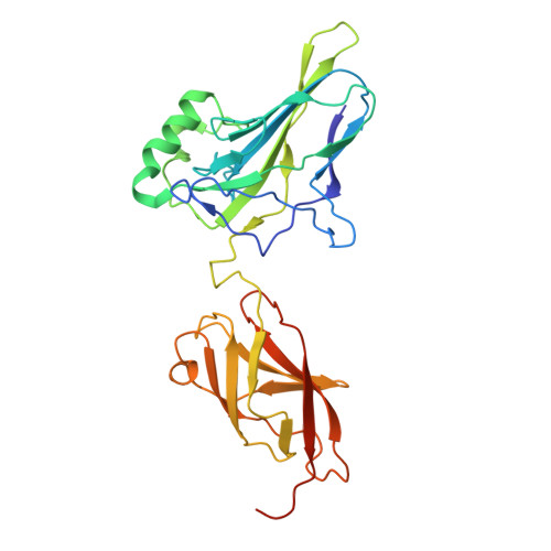
Teamwork of clustered low-affinity kappa B sites and accessory factors regulates transcriptional strength of NF-kappa B RelA dimers.
Shahabi, S., Biswas, T., Shen, Y., Sanahmadi, R., Zou, Y., Ghosh, G.(2025) Nucleic Acids Res 53
- PubMed: 41063342 Search on PubMedSearch on PubMed Central
- DOI: https://doi.org/10.1093/nar/gkaf846
- Primary Citation Related Structures:
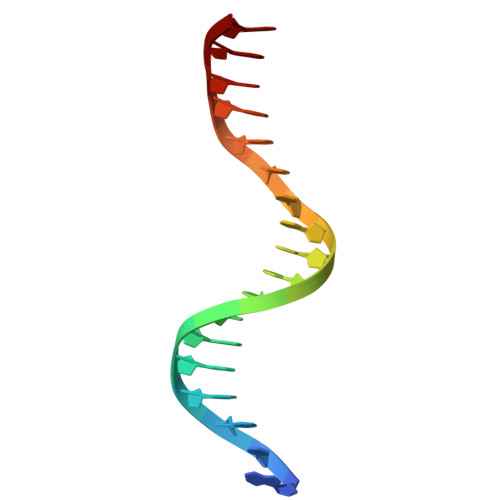
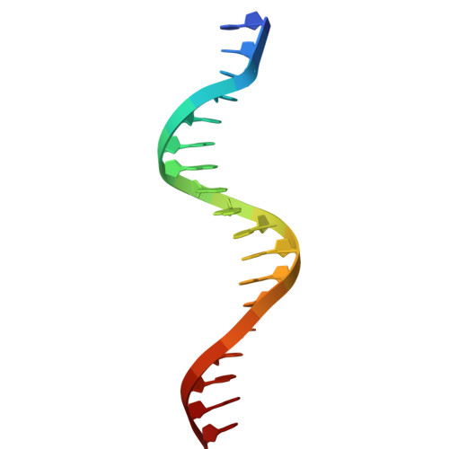
9E6W - PubMed Abstract:
Non-consensus binding sites of transcription factors (TFs) are often observed within the regulatory elements of genes; however, their effect on transcriptional strength is unclear. Within the promoters and enhancers of NF-κB-responsive genes, we identified clusters of non-consensus κB DNA sites, many exhibiting low affinity for NF-κB in vitro. Deletion of these sites demonstrated their collective critical role in transcription. We explored how these "weak" κB sites exert their influence, especially given the typically low nuclear concentrations of NF-κB. Using proteomics approaches, we identified additional nuclear factors, including other DNA-binding TFs, that could interact with κB site-bound NF-κB RelA. ChIP-seq and RNA-seq analyses suggest that these accessory TFs, referred to as the TF-cofactors of NF-κB, facilitate dynamic recruitment of NF-κB to the clustered weak κB sites. Overall, the occupancy of NF-κB at promoters and enhancers appears to be defined by a collective contribution from all κB sites, both weak and strong, in association with specific cofactors. This congregation of multiple factors within dynamic transcriptional complexes is likely a common feature of transcriptional programs.
- Department of Chemistry and Biochemistry, University of California San Diego, 9500 Gilman Drive, La Jolla,CA 92093, United States.
Organizational Affiliation: