A polycyclic scaffold identified by structure-based drug design effectively inhibits the human P2X7 receptor.
Oken, A.C., Turcu, A.L., Tzortzini, E., Georgiou, K., Nagel, J., Westermann, F.G., Barniol-Xicota, M., Seidler, J., Kim, G.R., Lee, S.D., Nicke, A., Kim, Y.C., Muller, C.E., Kolocouris, A., Vazquez, S., Mansoor, S.E.(2025) Nat Commun 16: 8283-8283
- PubMed: 40954149 Search on PubMedSearch on PubMed Central
- DOI: https://doi.org/10.1038/s41467-025-62643-8
- Primary Citation Related Structures:
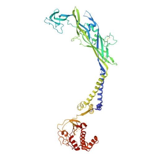
9E3M, 9E3N, 9E3O, 9E3P, 9E3Q - PubMed Abstract:
The P2X7 receptor is an ATP-gated ion channel that activates inflammatory pathways involved in diseases such as cancer, atherosclerosis, and neurodegeneration. However, despite the potential benefits of blocking overactive signaling, no P2X7 receptor antagonists have been approved for clinical use. Understanding species-specific pharmacological effects of existing antagonists has been challenging, in part due to the dearth of molecular information on receptor orthologs. Here, to identify distinct molecular features in the human receptor, we determine high-resolution cryo-EM structures of the full-length wild-type human P2X7 receptor in apo closed and ATP-bound open state conformations and draw comparisons with structures of other orthologs. We also report a cryo-EM structure of the human receptor in complex with an adamantane-based inhibitor, which we leverage, in conjunction with functional data and molecular dynamics simulations, to design a potent and selective antagonist with a unique polycyclic scaffold. Functional and structural analysis reveal how this optimized ligand, termed UB-MBX-46, interacts with the classical allosteric pocket of the human P2X7 receptor with subnanomolar potency and high selectivity, revealing its significant therapeutic potential.
- Department of Chemical Physiology & Biochemistry, Oregon Health & Science University, Portland, OR, USA.
Organizational Affiliation: