9DXX | pdb_00009dxx

Crystal structure of the A/Puerto Rico/8/1934 (H1N1) influenza virus hemagglutinin in complex with D-peptide


Experimental Data Snapshot

  • Method: X-RAY DIFFRACTION
  • Resolution: 2.37 Å
  • R-Value Free: 
    0.239 (Depositor), 0.244 (DCC) 
  • R-Value Work: 
    0.195 (Depositor), 0.199 (DCC) 
  • R-Value Observed: 
    0.197 (Depositor) 

Starting Model: experimental
View more details

wwPDB Validation   3D Report Full Report


Ligand Structure Quality Assessment 


This is version 2.0 of the entry. See complete history


Literature

De novo design of D-peptide ligands: Application to influenza virus hemagglutinin.

Juraszek, J.Kadam, R.U.Branduardi, D.van Ameijde, J.Garg, D.Dailly, N.Jongeneelen, M.Vermond, J.Brakenhoff, J.P.J.Brandenburg, B.van Dongen, M.J.P.Vogels, R.Friesen, R.H.E.Wilson, I.A.

(2025) Proc Natl Acad Sci U S A 122: e2426554122-e2426554122

  • DOI: https://doi.org/10.1073/pnas.2426554122
  • Primary Citation of Related Structures:  
    9DXX

  • PubMed Abstract: 

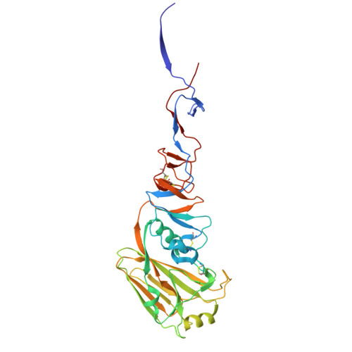
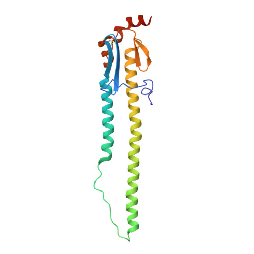
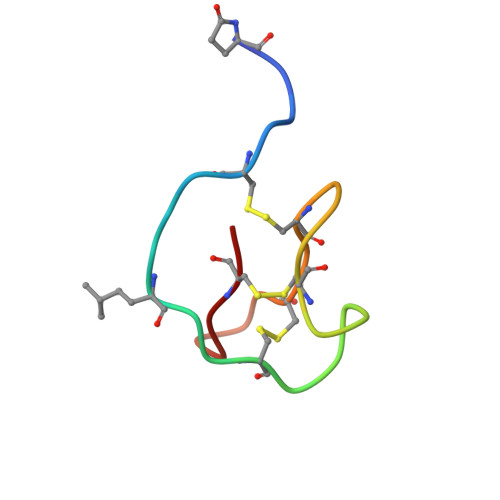
    D-peptides hold great promise as therapeutics by alleviating the challenges of metabolic stability and immunogenicity in L-peptides. However, current D-peptide discovery methods are severely limited by specific size, structure, and the chemical synthesizability of their protein targets. Here, we describe a computational method for de novo design of D-peptides that bind to an epitope of interest on the target protein using Rosetta's hotspot-centric approach. The approach comprises identifying hotspot sidechains in a functional protein-protein interaction and grafting these side chains onto much smaller structured peptide scaffolds of opposite chirality. The approach enables more facile design of D-peptides and its applicability is demonstrated by design of D-peptidic binders of influenza A virus hemagglutinin, resulting in identification of multiple D-peptide lead series. The X-ray structure of one of the leads at 2.38 Å resolution verifies the validity of the approach. This method should be generally applicable to targets with detailed structural information, independent of molecular size, and accelerate development of stable, peptide-based therapeutics.


  • Organizational Affiliation
    • Johnson and Johnson Innovative Medicine, Leiden 2333 CN, The Netherlands.

Macromolecules
Find similar proteins by:  (by identity cutoff)  |  3D Structure
Entity ID: 1
MoleculeChains Sequence LengthOrganismDetailsImage
Hemagglutinin HA1 chain328Influenza A virus (A/Puerto Rico/8/1934(H1N1))Mutation(s): 0 
Gene Names: HA
UniProt
Find proteins for P03452 (Influenza A virus (strain A/Puerto Rico/8/1934 H1N1))
Explore P03452 
Go to UniProtKB:  P03452
Entity Groups  
Sequence Clusters30% Identity50% Identity70% Identity90% Identity95% Identity100% Identity
UniProt GroupP03452
Glycosylation
Glycosylation Sites: 3
Sequence Annotations
Expand
  • Reference Sequence
Find similar proteins by:  (by identity cutoff)  |  3D Structure
Entity ID: 2
MoleculeChains Sequence LengthOrganismDetailsImage
Hemagglutinin HA2 chain176Influenza A virus (A/Puerto Rico/8/1934(H1N1))Mutation(s): 0 
Gene Names: HA
UniProt
Find proteins for P03452 (Influenza A virus (strain A/Puerto Rico/8/1934 H1N1))
Explore P03452 
Go to UniProtKB:  P03452
Entity Groups  
Sequence Clusters30% Identity50% Identity70% Identity90% Identity95% Identity100% Identity
UniProt GroupP03452
Glycosylation
Glycosylation Sites: 1
Sequence Annotations
Expand
  • Reference Sequence
Find similar proteins by:  (by identity cutoff)  |  3D Structure
Entity ID: 3
MoleculeChains Sequence LengthOrganismDetailsImage
D-peptideC [auth E]31Phage display vector pdvec0Mutation(s): 0 
Sequence Annotations
Expand
  • Reference Sequence
Oligosaccharides

Help

Entity ID: 4
MoleculeChains Length2D Diagram Glycosylation3D Interactions
alpha-D-mannopyranose-(1-3)-[alpha-D-mannopyranose-(1-6)]beta-D-mannopyranose-(1-4)-2-acetamido-2-deoxy-beta-D-glucopyranose-(1-4)-[alpha-L-fucopyranose-(1-3)][alpha-L-fucopyranose-(1-6)]2-acetamido-2-deoxy-beta-D-glucopyranoseD [auth L]7N-Glycosylation
Glycosylation Resources
GlyTouCan:  G11460AB
GlyCosmos:  G11460AB
GlyGen:  G11460AB
Entity ID: 5
MoleculeChains Length2D Diagram Glycosylation3D Interactions
2-acetamido-2-deoxy-beta-D-glucopyranose-(1-4)-2-acetamido-2-deoxy-beta-D-glucopyranoseE [auth C],
F [auth D]
2N-Glycosylation
Glycosylation Resources
GlyTouCan:  G42666HT
GlyCosmos:  G42666HT
GlyGen:  G42666HT
Small Molecules
Modified Residues  2 Unique
IDChains TypeFormula2D DiagramParent
7YO
Query on 7YO
C [auth E]PEPTIDE LINKINGC5 H7 N O3PRO
KW4
Query on KW4
C [auth E]D-PEPTIDE LINKINGC7 H15 N O2DLE
Experimental Data & Validation

Experimental Data

  • Method: X-RAY DIFFRACTION
  • Resolution: 2.37 Å
  • R-Value Free:  0.239 (Depositor), 0.244 (DCC) 
  • R-Value Work:  0.195 (Depositor), 0.199 (DCC) 
  • R-Value Observed: 0.197 (Depositor) 
Space Group: H 3 2
Unit Cell:
Length ( Å )Angle ( ˚ )
a = 107.693α = 90
b = 107.693β = 90
c = 387.162γ = 120
Software Package:
Software NamePurpose
PHENIXrefinement
HKL-2000data reduction
HKL-2000data scaling
PHASERphasing

Structure Validation

View Full Validation Report


Ligand Structure Quality Assessment 


Entry History & Funding Information

Deposition Data


Funding OrganizationLocationGrant Number
National Institutes of Health/National Institute Of Allergy and Infectious Diseases (NIH/NIAID)United StatesR56 AI117675

Revision History  (Full details and data files)

  • Version 1.0: 2025-06-25
    Type: Initial release
  • Version 1.1: 2025-07-16
    Changes: Database references
  • Version 1.2: 2025-07-23
    Changes: Database references
  • Version 2.0: 2025-07-30
    Changes: Atomic model, Database references, Derived calculations