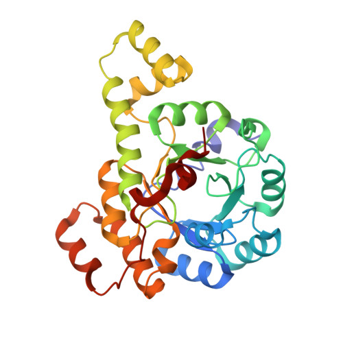
Structure and identification of the native PLP synthase complex from Methanosarcina acetivorans lysate.
Agnew, A., Humm, E., Zhou, K., Gunsalus, R.P., Zhou, Z.H.(2025) mBio 16: e0309024-e0309024
- PubMed: 39589128 Search on PubMed
- DOI: https://doi.org/10.1128/mbio.03090-24
- Primary Citation Related Structures:
9DVF - PubMed Abstract:
Many protein-protein interactions behave differently in biochemically purified forms as compared to their in vivo states. As such, determining native protein structures may elucidate structural states previously unknown for even well-characterized proteins. Here, we apply the bottom-up structural proteomics method, cryoID , toward a model methanogenic archaeon. While they are keystone organisms in the global carbon cycle and active members of the human microbiome, there is a general lack of characterization of methanogen enzyme structure and function. Through the cryoID approach, we successfully reconstructed and identified the native Methanosarcina acetivorans pyridoxal 5'-phosphate (PLP) synthase (PdxS) complex directly from cryogenic electron microscopy (cryo-EM) images of fractionated cellular lysate. We found that the native PdxS complex exists as a homo-dodecamer of PdxS subunits, and the previously proposed supracomplex containing both the synthase (PdxS) and glutaminase (PdxT) was not observed in cellular lysate. Our structure shows that the native PdxS monomer fashions a single 8α/8β TIM-barrel domain, surrounded by seven additional helices to mediate solvent and interface contacts. A density is present at the active site in the cryo-EM map and is interpreted as ribose 5-phosphate. In addition to being the first reconstruction of the PdxS enzyme from a heterogeneous cellular sample, our results reveal a departure from previously published archaeal PdxS crystal structures, lacking the 37-amino-acid insertion present in these prior cases. This study demonstrates the potential of applying the cryoID workflow to capture native structural states at atomic resolution for archaeal systems, for which traditional biochemical sample preparation is nontrivial.IMPORTANCEArchaea are one of the three domains of life, classified as a phylogenetically distinct lineage. There is a paucity of known enzyme structures from organisms of this domain, and this is often exacerbated by characteristically difficult growth conditions and a lack of readily available molecular biology toolkits to study proteins in archaeal cells. As a result, there is a gap in knowledge concerning the mechanisms governing archaeal protein behavior and their impacts on both the environment and human health; case in point, the synthesis of the widely utilized cofactor pyridoxal 5'-phosphate (PLP; a vitamer of vitamin B6, which humans cannot produce). By leveraging the power of single-particle cryo-EM and map-to-primary sequence identification, we determine the native structure of PLP synthase from cellular lysate. Our workflow allows the (i) rapid examination of new or less characterized systems with minimal sample requirements and (ii) discovery of structural states inaccessible by recombinant expression.
- Department of Microbiology, Immunology & Molecular Genetics, University of California Los Angeles, Los Angeles, California, USA.
Organizational Affiliation: