9DQX | pdb_00009dqx

Structure of western equine encephalitis virus CBA87 VLP


Experimental Data Snapshot

  • Method: ELECTRON MICROSCOPY
  • Resolution: 3.40 Å
  • Aggregation State: PARTICLE 
  • Reconstruction Method: SINGLE PARTICLE 

wwPDB Validation   3D Report Full Report


This is version 1.2 of the entry. See complete history


Literature

Molecular basis for shifted receptor recognition by an encephalitic arbovirus.

Fan, X.Li, W.Oros, J.Plante, J.A.Mitchell, B.M.Plung, J.S.Basu, H.Nagappan-Chettiar, S.Boeckers, J.M.Tjang, L.V.Mann, C.J.Brusic, V.Buck, T.K.Varnum, H.Yang, P.Malcolm, L.M.Choi, S.Y.de Souza, W.M.Chiu, I.M.Umemori, H.Weaver, S.C.Plante, K.S.Abraham, J.

(2025) Cell 188: 2957-2973.e28

  • DOI: https://doi.org/10.1016/j.cell.2025.03.029
  • Primary Citation Related Structures: 
    9DQV, 9DQX, 9DQY, 9DQZ

  • PubMed Abstract: 

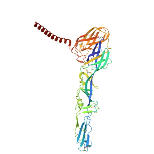
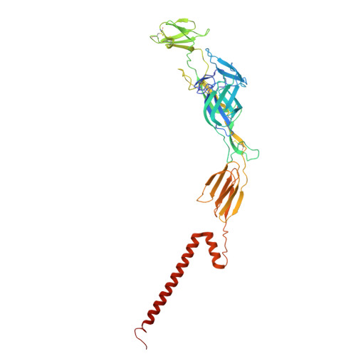
    Western equine encephalitis virus (WEEV) is an arbovirus that historically caused large outbreaks of encephalitis throughout the Americas. WEEV binds protocadherin 10 (PCDH10) as a receptor, and highly virulent ancestral WEEV strains also bind low-density lipoprotein receptor (LDLR)-related proteins. As WEEV declined as a human pathogen in North America over the past century, isolates have lost the ability to bind mammalian receptors while still recognizing avian receptors. To explain shifts in receptor dependencies and assess the risk of WEEV re-emergence, we determined cryoelectron microscopy structures of WEEV bound to human PCDH10, avian PCDH10, and human very-low-density lipoprotein receptor (VLDLR). We show that one to three E2 glycoprotein substitutions are sufficient for a nonpathogenic strain to regain the ability to bind mammalian receptors. A soluble VLDLR fragment protects mice from lethal challenge by a virulent ancestral WEEV strain. Because WEEV recently re-emerged in South America after decades of inactivity, our findings have important implications for outbreak preparedness.


  • Organizational Affiliation
    • Department of Microbiology, Blavatnik Institute, Harvard Medical School, Boston, MA, USA.

Macromolecules
Find similar proteins by:  (by identity cutoff)  |  3D Structure
Entity ID: 1
MoleculeChains Sequence LengthOrganismDetailsImage
Structural polyproteinA [auth J],
D,
E [auth G],
F [auth A]
437Western equine encephalitis virusMutation(s): 0 
UniProt
Find proteins for P13897 (Western equine encephalitis virus)
Explore P13897 
Go to UniProtKB:  P13897
Entity Groups  
Sequence Clusters30% Identity50% Identity70% Identity90% Identity95% Identity100% Identity
UniProt GroupP13897
Glycosylation
Glycosylation Sites: 1
Sequence Annotations
Expand
  • Reference Sequence
Find similar proteins by:  (by identity cutoff)  |  3D Structure
Entity ID: 2
MoleculeChains Sequence LengthOrganismDetailsImage
Structural polyproteinB [auth K],
G [auth E],
H,
I [auth B]
406Western equine encephalitis virusMutation(s): 0 
UniProt
Find proteins for P13897 (Western equine encephalitis virus)
Explore P13897 
Go to UniProtKB:  P13897
Entity Groups  
Sequence Clusters30% Identity50% Identity70% Identity90% Identity95% Identity100% Identity
UniProt GroupP13897
Glycosylation
Glycosylation Sites: 2
Sequence Annotations
Expand
  • Reference Sequence
Find similar proteins by:  (by identity cutoff)  |  3D Structure
Entity ID: 3
MoleculeChains Sequence LengthOrganismDetailsImage
Capsid proteinC [auth L],
J [auth F],
K [auth I],
L [auth C]
163Western equine encephalitis virusMutation(s): 0 
EC: 3.4.21.90
UniProt
Find proteins for P13897 (Western equine encephalitis virus)
Explore P13897 
Go to UniProtKB:  P13897
Entity Groups  
Sequence Clusters30% Identity50% Identity70% Identity90% Identity95% Identity100% Identity
UniProt GroupP13897
Sequence Annotations
Expand
  • Reference Sequence
Small Molecules
Ligands 1 Unique
IDChains Name / Formula / InChI Key2D Diagram3D Interactions
NAG (Subject of Investigation/LOI)
Query on NAG

Download Ideal Coordinates CCD File 
M [auth J]
N [auth K]
O [auth K]
P [auth D]
Q [auth G]
M [auth J],
N [auth K],
O [auth K],
P [auth D],
Q [auth G],
R [auth A],
S [auth E],
T [auth E],
U [auth H],
V [auth H],
W [auth B],
X [auth B]
2-acetamido-2-deoxy-beta-D-glucopyranose
C8 H15 N O6
OVRNDRQMDRJTHS-FMDGEEDCSA-N
Experimental Data & Validation

Experimental Data

  • Method: ELECTRON MICROSCOPY
  • Resolution: 3.40 Å
  • Aggregation State: PARTICLE 
  • Reconstruction Method: SINGLE PARTICLE 
EM Software:
TaskSoftware PackageVersion
MODEL REFINEMENTPHENIX

Structure Validation

View Full Validation Report


Entry History & Funding Information

Deposition Data


Funding OrganizationLocationGrant Number
National Institutes of Health/National Institute Of Allergy and Infectious Diseases (NIH/NIAID)United States--
Howard Hughes Medical Institute (HHMI)United States--

Revision History  (Full details and data files)

  • Version 1.0: 2025-03-26
    Type: Initial release
  • Version 1.1: 2025-05-28
    Changes: Data collection
  • Version 1.2: 2026-04-08
    Changes: Data collection, Database references