In vivo nucleotide excision repair by mycobacterial UvrD1 requires ATP hydrolysis but does not depend on cysteine disulfide-mediated dimerization and DNA unwinding.
Warren, G.M., Shuman, S.(2025) Nucleic Acids Res 53
- PubMed: 40193706 Search on PubMedSearch on PubMed Central
- DOI: https://doi.org/10.1093/nar/gkaf269
- Primary Citation Related Structures:
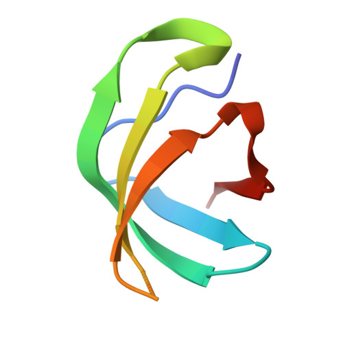
9DQS - PubMed Abstract:
Mycobacterial UvrD1 is an SF1-type ATPase that participates in nucleotide excision repair (NER). UvrD1 consists of N-terminal ATPase and C-terminal Tudor domains. The monomeric UvrD1 characterized originally displays vigorous DNA-dependent ATPase activity but only feeble helicase activity. A recent study demonstrated that: (i) cysteine disulfide-mediated homodimerization of UvrD1 generates a highly active helicase; and (ii) an obligate monomeric UvrD1 (by virtue of mutating the domain 2B cysteine) is active as an ATP-dependent 3'-to-5' single-stranded DNA translocase but not as a double-stranded DNA-unwinding helicase. Here we test genetically which physical and functional states of UvrD1 are relevant for its functions in DNA repair, by complementation of an NER-defective Mycobacterium smegmatis ΔuvrD1 strain with a series of biochemically-defined UvrD1 mutants. By assaying complemented strains for sensitivity to UVC, MMC, cisplatin, and psoralen-UVA, we conclude that monomeric UvrD1 ATPase activity suffices for the NER functions of UvrD1 in vivo. Decoupling ATP hydrolysis from duplex unwinding does not affect the repair activity of UvrD1, nor does interdiction of domain 2B cysteine disulfide-mediated dimerization or deletion of the Tudor domain. Our results militate against a proposed model in which UvrD1's repair function is governed by the redox state of the bacterium via its impact on UvrD1 dimerization and helicase activity.
- Molecular Biology Program, Memorial Sloan Kettering Cancer Center, NY, NY 10065, United States.
Organizational Affiliation: