9DOB | pdb_00009dob

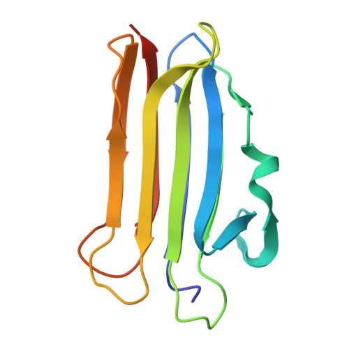
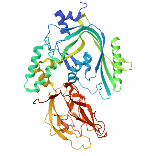
Membrane-bound OlyA (E69A)/PlyB (Hinge-Lock) prepore focused refinement


Experimental Data Snapshot

  • Method: ELECTRON MICROSCOPY
  • Resolution: 2.27 Å
  • Aggregation State: PARTICLE 
  • Reconstruction Method: SINGLE PARTICLE 

wwPDB Validation 3D Report Full Report

Validation slider image for 9DOB

This is version 1.1 of the entry. See complete history. 

Literature

Visualization of the interaction between sphingomyelin and cholesterol in lipid bilayer membranes.

Smothers, J.C., Nguyen, C.H.B., Han, Y., Li, Y., Chen, Z., Radhakrishnan, A.

(2026) Proc Natl Acad Sci U S A 123: e2528400123-e2528400123

  • DOI: https://doi.org/10.1073/pnas.2528400123
  • Primary Citation Related Structures: 
    9DO6, 9DO7, 9DO8, 9DO9, 9DOA, 9DOB

  • PubMed Abstract: 

    Biomembranes are complex two-dimensional liquids composed of hundreds of lipid species that interact in a myriad of ways. One such interaction, that between sphingomyelin (SM) and cholesterol in plasma membranes of animal cells, provides many functional benefits, including protection from microbial infection, prevention of unrestrained cell growth, and proper maintenance of cellular lipid composition. Owing to the liquid nature of membranes, the structure of the SM/cholesterol interaction, or any other functionally critical lipid-lipid interaction, has remained elusive. Here, we overcome this challenge using a fungal toxin called Ostreolysin A (OlyA), that has been shown to specifically bind to SM/cholesterol complexes in membranes. We used OlyA to stabilize the SM/cholesterol interaction much in the same way as antibodies are used to stabilize preexisting protein complexes. Cryoelectron microscopy analysis of OlyA bound to SM/cholesterol membranes reveals the details of the tight interaction between these two lipids-the steroid nucleus of cholesterol packs against the acyl chains of SM, and a hydrogen bond forms between the nitrogen on SM's ceramide base and the oxygen on cholesterol's hydroxyl group, thus sequestering this key functional group of cholesterol. The importance of hydrogen bonding in stabilizing the SM/cholesterol interaction is supported by structural analysis of a mutant form of OlyA that binds free SM in a cholesterol-independent manner. These results provide structural insights into the organization of cholesterol in membranes.


  • Organizational Affiliation: 
    • Department of Molecular Genetics, University of Texas, Southwestern Medical Center, Dallas, TX 75390-9046.

Macromolecule Content 

  • Total Structure Weight: 181.14 kDa 
  • Atom Count: 11,884 
  • Modeled Residue Count: 1,432 
  • Deposited Residue Count: 1,538 
  • Unique protein chains: 2

Macromolecules

Find similar proteins by:|  3D Structure
Entity ID: 1
MoleculeChains  Sequence LengthOrganismDetailsImage
Ostreolysin A6A,
B,
D [auth L],
E [auth M]
143Pleurotus ostreatusMutation(s): 1 
Gene Names: OlyA6
UniProt
Find proteins for P83467 (Pleurotus ostreatus)
Explore P83467 
Go to UniProtKB:  P83467
Entity Groups
Sequence Clusters30% Identity50% Identity70% Identity90% Identity95% Identity100% Identity
UniProt GroupP83467
Sequence Annotations
Expand
Reference Sequence
Find similar proteins by:|  3D Structure
Entity ID: 2
MoleculeChains  Sequence LengthOrganismDetailsImage
Pleurotolysin BC,
F [auth N]
483Pleurotus ostreatusMutation(s): 0 
Gene Names: plyB
UniProt
Find proteins for Q5W9E8 (Pleurotus ostreatus)
Explore Q5W9E8 
Go to UniProtKB:  Q5W9E8
Entity Groups
Sequence Clusters30% Identity50% Identity70% Identity90% Identity95% Identity100% Identity
UniProt GroupQ5W9E8
Sequence Annotations
Expand
Reference Sequence

Small Molecules

Ligands 1 Unique
IDChains Name / Formula / InChI Key2D Diagram3D Interactions
A1A8R
(Subject of Investigation/LOI)

Query on A1A8R


Download:Ideal Coordinates CCD File
G [auth A]
H [auth A]
I [auth A]
J [auth A]
K [auth B]
G [auth A],
H [auth A],
I [auth A],
J [auth A],
K [auth B],
L [auth B],
M [auth B],
N [auth B],
O [auth L],
P [auth L],
Q [auth L],
R [auth L],
S [auth M],
T [auth M],
U [auth M],
V [auth M]
N-oleoyl-D-erythro-sphingosylphosphorylcholine
C41 H82 N2 O6 P
NBEADXWAAWCCDG-QDDWGVBQSA-O

Experimental Data & Validation

Experimental Data

  • Method: ELECTRON MICROSCOPY
  • Resolution: 2.27 Å
  • Aggregation State: PARTICLE 
  • Reconstruction Method: SINGLE PARTICLE 

Structure Validation

View Full Validation Report


Entry History 

& Funding Information

Deposition Data


Funding OrganizationLocationGrant Number
National Institutes of Health/National Heart, Lung, and Blood Institute (NIH/NHLBI)United StatesHL160487
National Institutes of Health/National Institute Of Allergy and Infectious Diseases (NIH/NIAID)United StatesAI158357
National Institutes of Health/National Institute of General Medical Sciences (NIH/NIGMS)United StatesT32GM131963
Welch FoundationUnited StatesI-1793
Leducq FoundationFrance19CVD04

Revision History  (Full details and data files)

  • Version 1.0: 2025-09-24
    Type: Initial release
  • Version 1.1: 2026-10-07
    Changes: Data collection, Database references