9DMX | pdb_00009dmx

Crystal structure of shark UrIg2 V-C1-C2-C3 domains


Experimental Data Snapshot

  • Method: X-RAY DIFFRACTION
  • Resolution: 2.81 Å
  • R-Value Free: 
    0.275 (Depositor), 0.277 (DCC) 
  • R-Value Work: 
    0.249 (Depositor), 0.250 (DCC) 
  • R-Value Observed: 
    0.250 (Depositor) 

Starting Models: experimental
View more details

wwPDB Validation 3D Report Full Report

Validation slider image for 9DMX

Ligand Structure Quality Assessment 


This is version 1.0 of the entry. See complete history

Literature

Origin of immunoglobulins and T cell receptors: A candidate gene for invasion by the RAG transposon.

Flajnik, M.F.Stanfield, R.L.Verissimo, A.Neely, H.R.Munoz-Merida, A.Criscitiello, M.F.Wilson, I.A.Ohta, Y.

(2025) Sci Adv 11: eadw1273-eadw1273

  • DOI: https://doi.org/10.1126/sciadv.adw1273
  • Primary Citation Related Structures: 
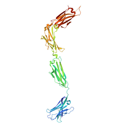
    9DMX

  • PubMed Abstract: 

    Rearranging antigen receptors (AgRs) arose when a variable (V) domain exon was invaded by the recombination-activating gene (RAG) transposon ~500 million years ago. We show here that the elasmobranch immunoglobulin heavy (IgH) isotypes-IgM, IgW, and IgNAR-are linked near the αδ T cell receptor (TCRαδ) locus. This linkage presages the emergence of the osteichthyan IgH translocon arrangement and clarifies the relationship between IgH and TCRδs. Recently, we reported UrIg , a nonrearranging, elasmobranch major histocompatibility complex (MHC)-linked AgR gene. Here, we describe a nonrearranging UrIg paralogue, UrIg2 , linked to this IgM/IgNAR/IgW/TCRαδ gene cluster in an AgR complex (AgRC). UrIg2 amino-terminal domains make homodimers where the C2-C3 structure resembles IgGFc. A relative of the UrIg2 V domain exon was invaded by the RAG transposon, revealing the genesis of the adaptive immune system. Our data indicate that an ancestral chromosome encoded an AgR precursor, undergoing RAG-mediated rearrangement after genome-wide duplication on one chromosome and retaining nonrearranging relics in the MHC and AgRC.


  • Organizational Affiliation
    • Department of Microbiology and Immunology, University of Maryland School of Medicine, Baltimore, MD 21201, USA.

Macromolecule Content 

  • Total Structure Weight: 104.21 kDa 
  • Atom Count: 6,969 
  • Modeled Residue Count: 869 
  • Deposited Residue Count: 914 
  • Unique protein chains: 1

Macromolecules

Find similar proteins by:|  3D Structure
Entity ID: 1
MoleculeChains  Sequence LengthOrganismDetailsImage
UrIg2 V-C1-C2-C3
A, B
457Ginglymostoma cirratumMutation(s): 0 

Oligosaccharides

Help  
Entity ID: 2
MoleculeChains Length2D Diagram GlycosylationD Interactions
beta-D-mannopyranose-(1-4)-2-acetamido-2-deoxy-beta-D-glucopyranose-(1-4)-[alpha-L-fucopyranose-(1-6)]2-acetamido-2-deoxy-beta-D-glucopyranose
C
4N-Glycosylation
Glycosylation Resources
GlyTouCan: G32152BH
GlyCosmos: G32152BH
GlyGen: G32152BH
Entity ID: 3
MoleculeChains Length2D Diagram GlycosylationD Interactions
2-acetamido-2-deoxy-beta-D-glucopyranose-(1-4)-2-acetamido-2-deoxy-beta-D-glucopyranose
D
2N-Glycosylation
Glycosylation Resources
GlyTouCan: G42666HT
GlyCosmos: G42666HT
GlyGen: G42666HT

Experimental Data & Validation

Experimental Data

  • Method: X-RAY DIFFRACTION
  • Resolution: 2.81 Å
  • R-Value Free:  0.275 (Depositor), 0.277 (DCC) 
  • R-Value Work:  0.249 (Depositor), 0.250 (DCC) 
  • R-Value Observed: 0.250 (Depositor) 
Space Group: P 21 21 21
Unit Cell:
Length ( Å )Angle ( ˚ )
a = 80.952α = 90
b = 97.384β = 90
c = 176.779γ = 90
Software Package:
Software NamePurpose
PHENIXrefinement
HKL-2000data reduction
HKL-2000data scaling
PHASERphasing

Structure Validation

View Full Validation Report


Ligand Structure Quality Assessment 


Entry History 

& Funding Information

Deposition Data


Funding OrganizationLocationGrant Number
Not funded--

Revision History  (Full details and data files)

  • Version 1.0: 2025-07-23
    Type: Initial release