Design of light- and chemically responsive protein assemblies through host-guest interactions.
Zhang, Z., Chiang, H.T., Xia, Y., Avakyan, N., Sonani, R.R., Wang, F., Egelman, E.H., De Yoreo, J.J., Pozzo, L.D., Tezcan, F.A.(2025) Chem 11
- PubMed: 40862094 Search on PubMedSearch on PubMed Central
- DOI: https://doi.org/10.1016/j.chempr.2024.102407
- Primary Citation Related Structures:
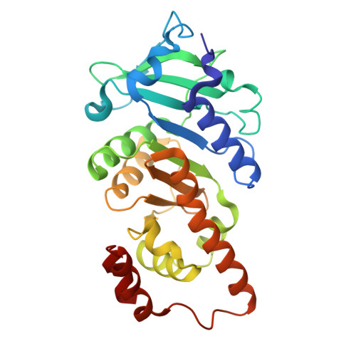
9DGH - PubMed Abstract:
Host-guest interactions have been widely used to build responsive materials and molecular machines owing to their inherently dynamic nature, interaction specificity, and responsiveness to diverse stimuli. Here we have set out to exploit these advantages of host-guest chemistry in the design of dynamic protein assemblies, using a C 4 symmetric protein, C98 RhuA, as a building block. We show that C98 RhuA variant individually modified with β-cyclodextrin (βCD) (host) or azobenzene (guest) functionalities can specifically pair with each other to form highly ordered 1- and 2-D assemblies. Association and dissociation βCD RhuA- azo RhuA assemblies can be controlled by UV and visible light as well as by small-molecule modulators of βCD-azobenzene interactions. Kinetics analyses reveal that βCD RhuA- azo RhuA nanotubes assemble without a nucleation barrier, a highly unusual occurrence for helical supramolecular systems. Taken together, our findings provide a compelling example for achieving complex structural and dynamic outcomes in protein assembly through simple chemical design.
- Department of Chemistry and Biochemistry, University of California, San Diego, La Jolla, CA 92093, USA.
Organizational Affiliation: