9DCA | pdb_00009dca

S. thermophilus class III ribonucleotide reductase with ATP and TTP

  • Classification: OXIDOREDUCTASE
  • Organism(s): Streptococcus thermophilus
  • Expression System: Escherichia coli
  • Mutation(s): No 

  • Deposited: 2024-08-25 Released: 2025-12-17 
  • Deposition Author(s): Andree, G.A., Drennan, C.L.
  • Funding Organization(s): National Institutes of Health/National Institute of General Medical Sciences (NIH/NIGMS), National Institutes of Health/National Institute of Environmental Health Sciences (NIH/NIEHS), Howard Hughes Medical Institute (HHMI)

Experimental Data Snapshot

  • Method: ELECTRON MICROSCOPY
  • Resolution: 3.60 Å
  • Aggregation State: PARTICLE 
  • Reconstruction Method: SINGLE PARTICLE 

wwPDB Validation 3D Report Full Report

Validation slider image for 9DCA

This is version 1.1 of the entry. See complete history

Literature

How ATP and dATP reposition class III ribonucleotide reductase cone domains to regulate enzyme activity.

Andree, G.A.Miller-Brown, K.R.Zhao, Z.Smith, A.K.Dawson, C.D.Deredge, D.J.Drennan, C.L.

(2025) Sci Adv 11: eady9156-eady9156

  • DOI: https://doi.org/10.1126/sciadv.ady9156
  • Primary Citation Related Structures: 
    9D3X, 9DAU, 9DCA

  • PubMed Abstract: 

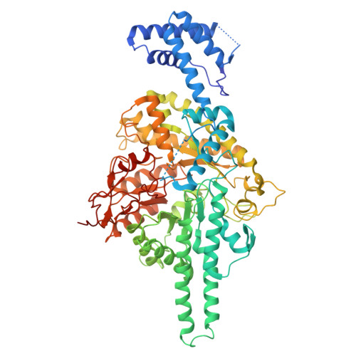
    Ribonucleotide reductases (RNRs) catalyze the conversion of ribonucleotides to deoxyribonucleotides. In the majority of cases, RNR activity is allosterically regulated by the cellular 2'-deoxyadenosine 5'-triphosphate (dATP)/adenosine 5'-triphosphate (ATP) ratio. To investigate allosteric activity regulation in anaerobic or class III (glycyl radical containing) RNRs, we determine cryo-electron microscopy structures of the class III RNR from Streptococcus thermophilus (StNrdD). We find that StNrdD's regulatory "cone" domains adopt markedly different conformations depending on whether the activator ATP or the inhibitor dATP is bound and that these different conformations alternatively position an "active site flap" toward the active site (ATP-bound) or away (dATP-bound). In contrast, the position of the glycyl radical domain is unaffected by the cone domain conformations, suggesting that StNrdD activity is regulated through control of substrate binding rather than control of radical transfer. Hydrogen-deuterium exchange mass spectrometry and mutagenesis support the structural findings. In addition, our structural data provide insight into the molecular basis by which ATP and dATP binding lead to the observed differential cone domain conformations.


  • Organizational Affiliation
    • Department of Chemistry, Massachusetts Institute of Technology, Cambridge, MA 02139, USA.

Macromolecule Content 

  • Total Structure Weight: 170.28 kDa 
  • Atom Count: 11,376 
  • Modeled Residue Count: 1,398 
  • Deposited Residue Count: 1,468 
  • Unique protein chains: 1

Macromolecules

Find similar proteins by:|  3D Structure
Entity ID: 1
MoleculeChains  Sequence LengthOrganismDetailsImage
Anaerobic ribonucleoside-triphosphate reductase
A, B
734Streptococcus thermophilusMutation(s): 0 
Gene Names: DF198_09700DQL92_10155
EC: 1.17.4.2
UniProt
Find proteins for A0A3G6JS83 (Streptococcus thermophilus)
Explore A0A3G6JS83 
Go to UniProtKB:  A0A3G6JS83
Entity Groups
Sequence Clusters30% Identity50% Identity70% Identity90% Identity95% Identity100% Identity
UniProt GroupA0A3G6JS83
Sequence Annotations
Expand
Reference Sequence

Small Molecules

Ligands 4 Unique
IDChains Name / Formula / InChI Key2D Diagram3D Interactions
ATP
(Subject of Investigation/LOI)

Query on ATP


Download:Ideal Coordinates CCD File
C [auth A],
D [auth A],
I [auth B],
J [auth B]
ADENOSINE-5'-TRIPHOSPHATE
C10 H16 N5 O13 P3
ZKHQWZAMYRWXGA-KQYNXXCUSA-N
TTP
(Subject of Investigation/LOI)

Query on TTP


Download:Ideal Coordinates CCD File
G [auth A],
H [auth B]
THYMIDINE-5'-TRIPHOSPHATE
C10 H17 N2 O14 P3
NHVNXKFIZYSCEB-XLPZGREQSA-N
ZN

Query on ZN


Download:Ideal Coordinates CCD File
M [auth B]ZINC ION
Zn
PTFCDOFLOPIGGS-UHFFFAOYSA-N
MG

Query on MG


Download:Ideal Coordinates CCD File
E [auth A],
F [auth A],
K [auth B],
L [auth B]
MAGNESIUM ION
Mg
JLVVSXFLKOJNIY-UHFFFAOYSA-N

Experimental Data & Validation

Experimental Data

  • Method: ELECTRON MICROSCOPY
  • Resolution: 3.60 Å
  • Aggregation State: PARTICLE 
  • Reconstruction Method: SINGLE PARTICLE 
EM Software:
TaskSoftware PackageVersion
MODEL REFINEMENTPHENIX1.21.1_5286
RECONSTRUCTIONRELION4.0.0

Structure Validation

View Full Validation Report


Entry History 

& Funding Information

Deposition Data


Funding OrganizationLocationGrant Number
National Institutes of Health/National Institute of General Medical Sciences (NIH/NIGMS)United StatesGM126982
National Institutes of Health/National Institute of Environmental Health Sciences (NIH/NIEHS)United StatesT32ES007020
Howard Hughes Medical Institute (HHMI)United States--

Revision History  (Full details and data files)

  • Version 1.0: 2025-12-17
    Type: Initial release
  • Version 1.1: 2025-12-24
    Changes: Data collection, Database references