9D9O | pdb_00009d9o

Crystal structure of CVB3 replication-linked RNA stem-loop D in complex with 3C protein


Experimental Data Snapshot

  • Method: X-RAY DIFFRACTION
  • Resolution: 2.41 Å
  • R-Value Free: 
    0.238 (Depositor), 0.238 (DCC) 
  • R-Value Work: 
    0.194 (Depositor), 0.195 (DCC) 
  • R-Value Observed: 
    0.199 (Depositor) 

Starting Model: experimental
View more details

wwPDB Validation   3D Report Full Report


This is version 1.1 of the entry. See complete history


Literature

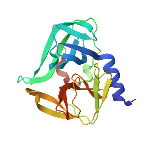
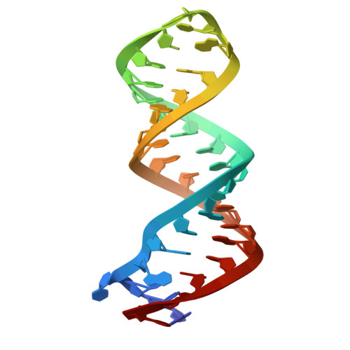
Structural basis for 3C and 3CD recruitment by enteroviral genomes during negative-strand RNA synthesis.

Das, N.K.Patel, A.Abdelghani, R.Koirala, D.

(2025) Nat Commun 16: 9293-9293

  • DOI: https://doi.org/10.1038/s41467-025-64376-0
  • Primary Citation Related Structures: 
    9D9O, 9D9P

  • PubMed Abstract: 

    Enteroviral replication-linked cloverleaf RNAs recruit the viral 3CD protein, a fusion of 3C protease and 3D RNA-dependent RNA-polymerase, for negative-strand synthesis during genome replication. However, the structures and mechanisms of this virological process remain unclear. Using the coxsackievirus B3 model, we determine the crystal structures of both intact cloverleaf-3C and isolated sD-3C complexes at 2.69 Å and 2.41 Å resolutions, respectively. Our structures reveal that the sD stem-loop is the sole determinant for binding two 3C monomers, with each monomer recognizing the lateral surface of the sD stem either upstream (toward the apical tetraloop) or downstream (near the dinucleotide bulge) of the Py•Py helix. Binding studies with structure-guided cloverleaf and 3C mutants further clarify the roles of specific nucleotides and residues involved in the interactions between cloverleaf and 3C, explaining earlier virological observations. Through comparative structural and binding studies of 3C, 3D, and 3CD with cloverleafs from seven different enteroviral species, we demonstrate that while the 3D domain does not contribute to cloverleaf binding, the sD sequence and its structural pattern govern 3CD-cloverleaf interactions through the 3C domain. Our work establishes a high-resolution structural framework for understanding enteroviral replication mechanisms, which will aid in developing antivirals targeting this platform.


  • Organizational Affiliation
    • Department of Chemistry and Biochemistry, University of Maryland, Baltimore County (UMBC), Baltimore, MD, USA.

Macromolecules

Find similar proteins by:  (by identity cutoff)  |  3D Structure
Entity ID: 1
MoleculeChains Sequence LengthOrganismDetailsImage
Protease 3C
A, B
192Coxsackievirus B3Mutation(s): 1 
EC: 3.4.22.28
UniProt
Find proteins for P03313 (Coxsackievirus B3 (strain Nancy))
Explore P03313 
Go to UniProtKB:  P03313
Entity Groups  
Sequence Clusters30% Identity50% Identity70% Identity90% Identity95% Identity100% Identity
UniProt GroupP03313
Sequence Annotations
Expand
  • Reference Sequence
Find similar nucleic acids by:  (by identity cutoff) 
Entity ID: 2
MoleculeChains LengthOrganismImage
RNA (36-MER)C [auth R]36Coxsackievirus B3
Sequence Annotations
Expand
  • Reference Sequence
Experimental Data & Validation

Experimental Data

  • Method: X-RAY DIFFRACTION
  • Resolution: 2.41 Å
  • R-Value Free:  0.238 (Depositor), 0.238 (DCC) 
  • R-Value Work:  0.194 (Depositor), 0.195 (DCC) 
  • R-Value Observed: 0.199 (Depositor) 
Space Group: P 21 21 21
Unit Cell:
Length ( Å )Angle ( ˚ )
a = 52.3α = 90
b = 78.869β = 90
c = 106.508γ = 90
Software Package:
Software NamePurpose
PHENIXrefinement
PHENIXphasing
DIALSdata reduction
xia2data scaling
Cootmodel building

Structure Validation

View Full Validation Report


Entry History & Funding Information

Deposition Data


Funding OrganizationLocationGrant Number
National Science Foundation (NSF, United States)United StatesMCB-2236996

Revision History  (Full details and data files)

  • Version 1.0: 2025-10-01
    Type: Initial release
  • Version 1.1: 2026-04-15
    Changes: Database references