9CV8 | pdb_00009cv8

Crystal Structure of Cytochrome P450 NysL bound to nystatin


Experimental Data Snapshot

  • Method: X-RAY DIFFRACTION
  • Resolution: 2.00 Å
  • R-Value Free: 
    0.215 (Depositor), 0.217 (DCC) 
  • R-Value Work: 
    0.189 (Depositor), 0.191 (DCC) 
  • R-Value Observed: 
    0.190 (Depositor) 

Starting Model: experimental
View more details

wwPDB Validation   3D Report Full Report


Ligand Structure Quality Assessment 


This is version 1.1 of the entry. See complete history


Literature

Crystal structure of cytochrome P450 NysL and the structural basis for stereo- and regio-selective oxidation of antifungal macrolides.

Murarka, V.C.Kim, J.S.Lamb, D.C.Kelly, S.L.Poulos, T.L.Follmer, A.H.

(2025) J Biological Chem 301: 108185-108185

  • DOI: https://doi.org/10.1016/j.jbc.2025.108185
  • Primary Citation of Related Structures:  
    9CV8

  • PubMed Abstract: 

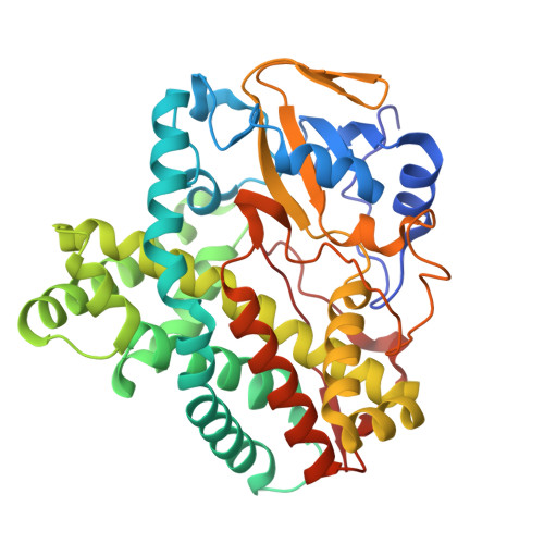
    NysL, a cytochrome P450 monooxygenase from the Gram-positive bacterium Streptomyces noursei, catalyzes the C10 hydroxylation of 10-deoxynystain to nystatin A 1 , a clinically important antifungal. In this study, we present the 2.0 Å resolution crystal structure of NysL bound to nystatin A 1 . The structure of this complex provides key insights into the structural elements that dictate the regio- and stereo- selective oxidation of large 20-44-membered macrolide substrates. The closely related AmphL operates on a similar 38-member macrolide but oxidizes C8 rather than C10. This difference requires that the substrate for AmphL penetrate further into the active site relative to NysL. The depth of substrate penetration is controlled by interactions between an area of the substrate binding pocket deemed the "back-wall" and the hemiketal ring of the macrolide substrate.


  • Organizational Affiliation
    • Department of Molecular Biology and Biochemistry, University of California, Irvine, California 92697-3900, United States. Electronic address: vmurarka@uci.edu.

Macromolecules
Find similar proteins by:  (by identity cutoff)  |  3D Structure
Entity ID: 1
MoleculeChains Sequence LengthOrganismDetailsImage
NysL
A, B
394Streptomyces nourseiMutation(s): 0 
Gene Names: nysL
UniProt
Find proteins for Q9L4X0 (Streptomyces noursei)
Explore Q9L4X0 
Go to UniProtKB:  Q9L4X0
Entity Groups  
Sequence Clusters30% Identity50% Identity70% Identity90% Identity95% Identity100% Identity
UniProt GroupQ9L4X0
Sequence Annotations
Expand
  • Reference Sequence
Small Molecules
Ligands 3 Unique
IDChains Name / Formula / InChI Key2D Diagram3D Interactions
A1AZ7 (Subject of Investigation/LOI)
Query on A1AZ7

Download Ideal Coordinates CCD File 
D [auth A],
K [auth B]
Nystatin
C47 H75 N O17
VQOXZBDYSJBXMA-NQTDYLQESA-N
HEM
Query on HEM

Download Ideal Coordinates CCD File 
C [auth A],
J [auth B]
PROTOPORPHYRIN IX CONTAINING FE
C34 H32 Fe N4 O4
KABFMIBPWCXCRK-RGGAHWMASA-L
SO4
Query on SO4

Download Ideal Coordinates CCD File 
E [auth A]
F [auth A]
G [auth A]
H [auth A]
I [auth A]
E [auth A],
F [auth A],
G [auth A],
H [auth A],
I [auth A],
L [auth B],
M [auth B],
N [auth B],
O [auth B]
SULFATE ION
O4 S
QAOWNCQODCNURD-UHFFFAOYSA-L
Experimental Data & Validation

Experimental Data

  • Method: X-RAY DIFFRACTION
  • Resolution: 2.00 Å
  • R-Value Free:  0.215 (Depositor), 0.217 (DCC) 
  • R-Value Work:  0.189 (Depositor), 0.191 (DCC) 
  • R-Value Observed: 0.190 (Depositor) 
Space Group: P 31 2 1
Unit Cell:
Length ( Å )Angle ( ˚ )
a = 136.29α = 90
b = 136.29β = 90
c = 135.6γ = 120
Software Package:
Software NamePurpose
PHENIXrefinement
Aimlessdata scaling
XDSdata reduction
PHASERphasing
PDB_EXTRACTdata extraction

Structure Validation

View Full Validation Report


Ligand Structure Quality Assessment 


Entry History & Funding Information

Deposition Data


Funding OrganizationLocationGrant Number
National Institutes of Health/National Institute of General Medical Sciences (NIH/NIGMS)United StatesGM131920

Revision History  (Full details and data files)

  • Version 1.0: 2025-01-29
    Type: Initial release
  • Version 1.1: 2025-02-26
    Changes: Database references