9CSN | pdb_00009csn

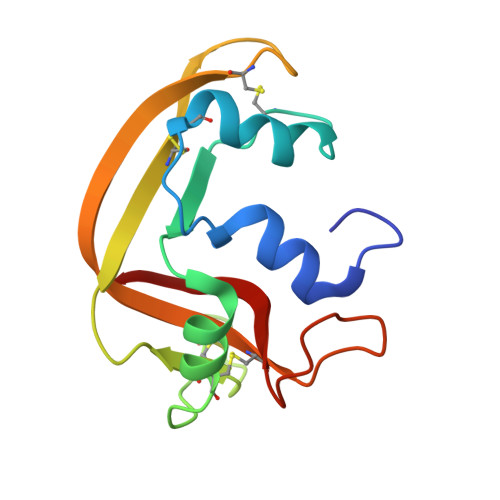
Crystal structure of human ribonuclease 7 (RNase 7) in complex with 5'-adenosine monophosphate (5'-AMP)

  • Classification: HYDROLASE
  • Organism(s): Homo sapiens
  • Expression System: Escherichia coli BL21(DE3)
  • Mutation(s): No 

  • Deposited: 2024-07-24 Released: 2025-07-30 
  • Deposition Author(s): Tran, T.T.Q., Pham, N.T.H., Calmettes, C., Doucet, N.
  • Funding Organization(s): Natural Sciences and Engineering Research Council (NSERC, Canada), Fonds de Recherche du Quebec - Sante (FRQS), Canada Foundation for Innovation, Fonds de Recherche du Quebec - Nature et Technologies (FRQNT)

Experimental Data Snapshot

  • Method: X-RAY DIFFRACTION
  • Resolution: 2.07 Å
  • R-Value Free: 
    0.262 (Depositor), 0.263 (DCC) 
  • R-Value Work: 
    0.217 (Depositor), 0.218 (DCC) 
  • R-Value Observed: 
    0.222 (Depositor) 

Starting Model: experimental
View more details

wwPDB Validation 3D Report Full Report

Validation slider image for 9CSN

Ligand Structure Quality Assessment 


This is version 1.1 of the entry. See complete history

Literature

Engineering human RNase 7 with an eosinophil RNase segment reveals determinants of cytotoxic and antimicrobial activity.

Tran, T.T.Q.Larda, S.T.Nieves, C.Veyrier, F.J.Calmettes, C.Doucet, N.

(2026) J Biol Chem 302: 113189-113189

  • DOI: https://doi.org/10.1016/j.jbc.2026.113189
  • Primary Citation Related Structures: 
    9CSM, 9CSN

  • PubMed Abstract: 

    Human ribonuclease 7 is a potent antimicrobial member of the RNase A superfamily with little cytotoxic activity, in contrast to the closely related eosinophil cationic protein (HsR3). To determine which structural elements underlie these divergent functions, we engineered a chimeric RNase 7 variant in which loop 4 to 5 residues 61 to 77 were replaced by the corresponding segment from HsR3. This chimera acquired cytotoxicity toward HeLa cells while retaining antibacterial activity against Escherichia coli and Mycobacterium smegmatis, identifying this segment as a key transferable determinant of RNase function. Consistent with this result, a peptide spanning HsR3 residues 61 to 77 also displayed antibacterial activity, supporting a direct contribution of this region to host-defense activity and functional modulation within the RNase scaffold. To define the structural basis of RNase 7 activity, we determined crystal structures of RNase 7 in apo and 5'-AMP-bound forms and combined these with NMR titrations and molecular docking. These analyses revealed a canonical RNase fold together with an unexpected adenine-binding pose in the B 1 subsite. Complementary binding analyses showed that RNase 7 preferentially recognizes pyrimidines, yet can also accommodate purine-containing ligands such as UpA in more than one binding mode. Together, these findings establish loop 4 to 5 as a portable functional module that can reprogram biological activity within the human RNase scaffold and provide a structural framework for engineering multifunctional RNases with tailored properties.


  • Organizational Affiliation
    • Centre Armand-Frappier Santé Biotechnologie, Institut National de la Recherche Scientifique (INRS), Université du Québec, Laval, Quebec, Canada.

Macromolecule Content 

  • Total Structure Weight: 30.54 kDa 
  • Atom Count: 2,199 
  • Modeled Residue Count: 256 
  • Deposited Residue Count: 258 
  • Unique protein chains: 1

Macromolecules

Find similar proteins by:|  3D Structure
Entity ID: 1
MoleculeChains  Sequence LengthOrganismDetailsImage
Ribonuclease 7
A, B
129Homo sapiensMutation(s): 0 
Gene Names: RNASE7UNQ2516/PRO6006
EC: 3.1.27
UniProt & NIH Common Fund Data Resources
Find proteins for Q9H1E1 (Homo sapiens)
Explore Q9H1E1 
Go to UniProtKB:  Q9H1E1
PHAROS:  Q9H1E1
GTEx:  ENSG00000165799 
Entity Groups
Sequence Clusters30% Identity50% Identity70% Identity90% Identity95% Identity100% Identity
UniProt GroupQ9H1E1
Sequence Annotations
Expand
Reference Sequence

Small Molecules

Ligands 4 Unique
IDChains Name / Formula / InChI Key2D Diagram3D Interactions
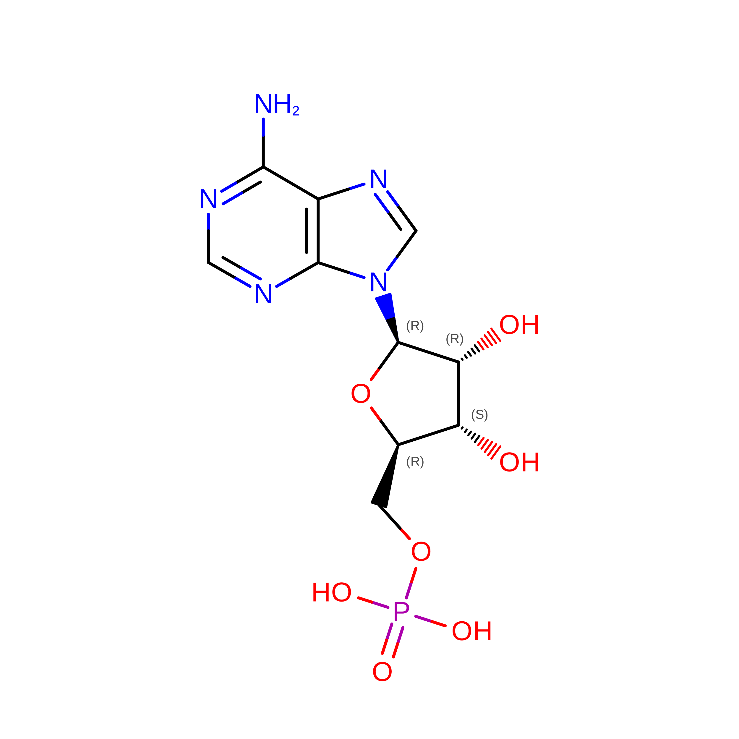
AMP
(Subject of Investigation/LOI)

Query on AMP


Download:Ideal Coordinates CCD File
F [auth A]ADENOSINE MONOPHOSPHATE
C10 H14 N5 O7 P
UDMBCSSLTHHNCD-KQYNXXCUSA-N
GOL

Query on GOL


Download:Ideal Coordinates CCD File
G [auth A],
P [auth B]
GLYCEROL
C3 H8 O3
PEDCQBHIVMGVHV-UHFFFAOYSA-N
EDO

Query on EDO


Download:Ideal Coordinates CCD File
H [auth A],
K [auth A],
L [auth B],
M [auth B],
N [auth B]
1,2-ETHANEDIOL
C2 H6 O2
LYCAIKOWRPUZTN-UHFFFAOYSA-N
BO3

Query on BO3


Download:Ideal Coordinates CCD File
C [auth A]
D [auth A]
E [auth A]
I [auth A]
J [auth A]
C [auth A],
D [auth A],
E [auth A],
I [auth A],
J [auth A],
O [auth B]
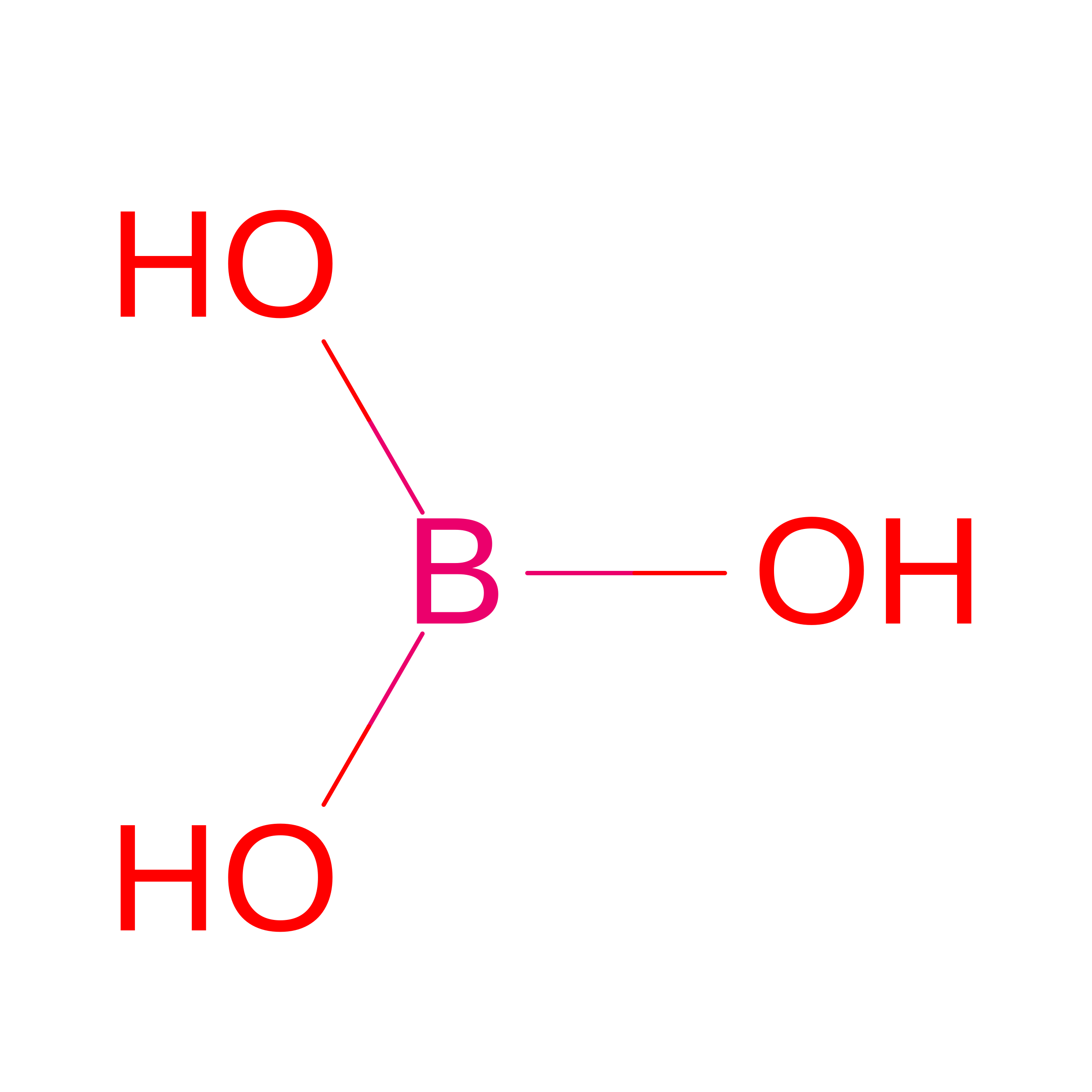
BORIC ACID
B H3 O3
KGBXLFKZBHKPEV-UHFFFAOYSA-N

Experimental Data & Validation

Experimental Data

  • Method: X-RAY DIFFRACTION
  • Resolution: 2.07 Å
  • R-Value Free:  0.262 (Depositor), 0.263 (DCC) 
  • R-Value Work:  0.217 (Depositor), 0.218 (DCC) 
  • R-Value Observed: 0.222 (Depositor) 
Space Group: P 1 21 1
Unit Cell:
Length ( Å )Angle ( ˚ )
a = 43.11α = 90
b = 48.17β = 97.28
c = 62.9γ = 90
Software Package:
Software NamePurpose
PHENIXrefinement
XSCALEdata scaling
PDB_EXTRACTdata extraction
PHASERphasing

Structure Validation

View Full Validation Report


Ligand Structure Quality Assessment 


Entry History 

& Funding Information

Deposition Data


Funding OrganizationLocationGrant Number
Natural Sciences and Engineering Research Council (NSERC, Canada)CanadaRGPIN-2022-04368
Natural Sciences and Engineering Research Council (NSERC, Canada)CanadaRGPIN-2017-06091
Natural Sciences and Engineering Research Council (NSERC, Canada)CanadaCREATE APRENTICE
Fonds de Recherche du Quebec - Sante (FRQS)Canada281993
Canada Foundation for InnovationCanadaMSI - GlycoNet Integrated Services
Fonds de Recherche du Quebec - Nature et Technologies (FRQNT)CanadaPROTEO

Revision History  (Full details and data files)

  • Version 1.0: 2025-07-30
    Type: Initial release
  • Version 1.1: 2026-08-12
    Changes: Database references