


Role of Electrostatics in Hydride Transfer by Horse liver alcohol dehydrogenase
Mukherjee, S., Fried, S.D.E., Boxer, S.G.To be published.
Experimental Data Snapshot
Starting Model: experimental
View more details
Entity ID: 1 | |||||
|---|---|---|---|---|---|
| Molecule | Chains | Sequence Length | Organism | Details | Image |
| Alcohol dehydrogenase E chain | 377 | Equus caballus | Mutation(s): 1 EC: 1.1.1.1 | 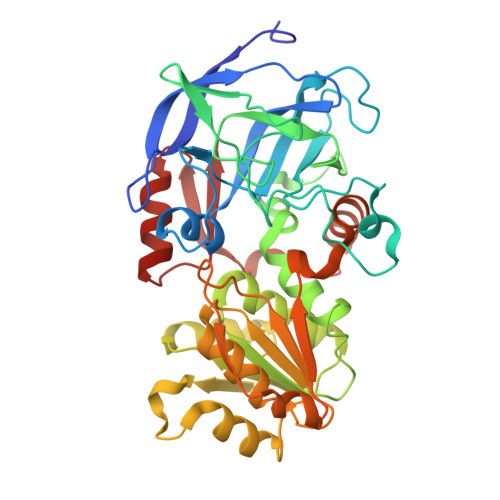 | |
UniProt | |||||
Entity Groups | |||||
| Sequence Clusters | 30% Identity50% Identity70% Identity90% Identity95% Identity100% Identity | ||||
| UniProt Group | P00327 | ||||
Sequence AnnotationsExpand | |||||
Reference Sequence | |||||
| Ligands 3 Unique | |||||
|---|---|---|---|---|---|
| ID | Chains | Name / Formula / InChI Key | 2D Diagram | 3D Interactions | |
| NAI (Subject of Investigation/LOI) Download:Ideal Coordinates CCD File | E [auth A], I [auth B] | 1,4-DIHYDRONICOTINAMIDE ADENINE DINUCLEOTIDE C21 H29 N7 O14 P2 BOPGDPNILDQYTO-NNYOXOHSSA-N |  | ||
| CXF (Subject of Investigation/LOI) Download:Ideal Coordinates CCD File | F [auth A], J [auth B] | CYCLOHEXYLFORMAMIDE C7 H13 N O SWGXDLRCJNEEGZ-UHFFFAOYSA-N |  | ||
| ZN (Subject of Investigation/LOI) Download:Ideal Coordinates CCD File | C [auth A], D [auth A], G [auth B], H [auth B] | ZINC ION Zn PTFCDOFLOPIGGS-UHFFFAOYSA-N |  | ||
| Length ( Å ) | Angle ( ˚ ) |
|---|---|
| a = 44.243 | α = 92.34 |
| b = 50.508 | β = 103.03 |
| c = 92.812 | γ = 109.22 |
| Software Name | Purpose |
|---|---|
| PHENIX | refinement |
| Aimless | data scaling |
| XDS | data reduction |
| PHASER | phasing |
| Funding Organization | Location | Grant Number |
|---|---|---|
| National Institutes of Health/Office of the Director | United States | R35GM118044 |


