9CJP | pdb_00009cjp

X-ray crystal structure of SARS-CoV-2 main protease quadruple mutants in complex with Nirmatrelvir


Experimental Data Snapshot

  • Method: X-RAY DIFFRACTION
  • Resolution: 1.71 Å
  • R-Value Free: 
    0.226 (Depositor), 0.226 (DCC) 
  • R-Value Work: 
    0.180 (Depositor), 0.180 (DCC) 
  • R-Value Observed: 
    0.182 (Depositor) 

Starting Model: experimental
View more details

wwPDB Validation   3D Report Full Report


Ligand Structure Quality Assessment 


This is version 1.1 of the entry. See complete history


Literature

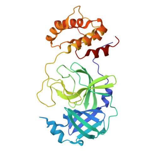
Structural basis for varying drug resistance of SARS-CoV-2 M pro E166 variants.

Esler, M.A.Shi, K.Rollie, J.A.Delgado, R.Vishwakarma, J.Dabrowska, A.Prahlad, J.Moghadasi, S.A.Harris, R.S.Aihara, H.

(2025) mBio 16: e0262424-e0262424

  • DOI: https://doi.org/10.1128/mbio.02624-24
  • Primary Citation of Related Structures:  
    9CJO, 9CJP, 9CJQ, 9CJR, 9CJS, 9CJT, 9CJU, 9CJV

  • PubMed Abstract: 

    Severe acute respiratory syndrome coronavirus 2 (SARS-CoV-2) main protease (M pro ) has an essential role in the virus lifecycle and, accordingly, it is a target for antiviral drugs. Multiple studies have identified an M pro mutation (E166V) that confers strong resistance to clinically relevant inhibitors, including nirmatrelvir, but the underlying mechanism is not fully understood. Here, we report on crystal structures of SARS-CoV-2 M pro E166V in complex with nirmatrelvir, ensitrelvir, and bofutrelvir. The structures suggest that resistance is caused in part by the loss of a direct hydrogen bond and also, especially for nirmatrelvir, by a steric clash with the substituted valine residue. In comparison, the binding of bofutrelvir shows greater flexibility, which may help alleviate this steric effect and allow bofutrelvir to fit the mutant active site despite the loss of a direct polar contact. Thermal stability analyses also corroborate E166V most severely affecting the binding of nirmatrelvir and, to lesser and different extents, ensitrelvir and bofutrelvir. We further show that E166I causes even more severe nirmatrelvir resistance, whereas E166A and E166L have much milder effects. These studies shed light on the molecular mechanisms of a key M pro drug resistance mutation and may help inform the design of next-generation inhibitors.IMPORTANCEUsing a combination of high-resolution X-ray crystallographic and biochemical analyses, we reveal the molecular mechanisms by which a mutation in the severe acute respiratory syndrome coronavirus 2 main protease (M pro ) confers strong resistance against clinically relevant antiviral drugs that inhibit M pro activity. The results presented here may help inform the design of next-generation inhibitors to combat the problem of therapy resistance.


  • Organizational Affiliation
    • Department of Biochemistry, Molecular Biology and Biophysics, University of Minnesota, Minneapolis, Minnesota, USA.

Macromolecules
Find similar proteins by:  (by identity cutoff)  |  3D Structure
Entity ID: 1
MoleculeChains Sequence LengthOrganismDetailsImage
3C-like proteinase nsp5
A, B
306Severe acute respiratory syndrome coronavirus 2Mutation(s): 4 
Gene Names: rep1a-1b
EC: 3.4.22.69
UniProt
Find proteins for P0DTD1 (Severe acute respiratory syndrome coronavirus 2)
Explore P0DTD1 
Go to UniProtKB:  P0DTD1
Entity Groups  
Sequence Clusters30% Identity50% Identity70% Identity90% Identity95% Identity100% Identity
UniProt GroupP0DTD1
Sequence Annotations
Expand
  • Reference Sequence
Small Molecules
Ligands 4 Unique
IDChains Name / Formula / InChI Key2D Diagram3D Interactions
4WI (Subject of Investigation/LOI)
Query on 4WI

Download Ideal Coordinates CCD File 
C [auth A],
F [auth B]
(1R,2S,5S)-N-{(1E,2S)-1-imino-3-[(3S)-2-oxopyrrolidin-3-yl]propan-2-yl}-6,6-dimethyl-3-[3-methyl-N-(trifluoroacetyl)-L-valyl]-3-azabicyclo[3.1.0]hexane-2-carboxamide
C23 H34 F3 N5 O4
WDVIRQQKRMIXGS-XIFHJVQQSA-N
SO4
Query on SO4

Download Ideal Coordinates CCD File 
J [auth B]SULFATE ION
O4 S
QAOWNCQODCNURD-UHFFFAOYSA-L
BR
Query on BR

Download Ideal Coordinates CCD File 
E [auth A],
H [auth B],
I [auth B]
BROMIDE ION
Br
CPELXLSAUQHCOX-UHFFFAOYSA-M
EDO
Query on EDO

Download Ideal Coordinates CCD File 
D [auth A],
G [auth B]
1,2-ETHANEDIOL
C2 H6 O2
LYCAIKOWRPUZTN-UHFFFAOYSA-N
Experimental Data & Validation

Experimental Data

  • Method: X-RAY DIFFRACTION
  • Resolution: 1.71 Å
  • R-Value Free:  0.226 (Depositor), 0.226 (DCC) 
  • R-Value Work:  0.180 (Depositor), 0.180 (DCC) 
  • R-Value Observed: 0.182 (Depositor) 
Space Group: P 1 21 1
Unit Cell:
Length ( Å )Angle ( ˚ )
a = 48.095α = 90
b = 105.665β = 103.61
c = 53.1γ = 90
Software Package:
Software NamePurpose
PHENIXrefinement
XDSdata reduction
STARANISOdata scaling
PHASERphasing

Structure Validation

View Full Validation Report


Ligand Structure Quality Assessment 


Entry History & Funding Information

Deposition Data


Funding OrganizationLocationGrant Number
National Institutes of Health/National Cancer Institute (NIH/NCI)United StatesP01 CA214279
National Institutes of Health/National Institute Of Allergy and Infectious Diseases (NIH/NIAID)United StatesR37 AI064064

Revision History  (Full details and data files)

  • Version 1.0: 2025-07-09
    Type: Initial release
  • Version 1.1: 2026-01-21
    Changes: Database references