9CGH | pdb_00009cgh

Photoactive yellow protein crystallized in situ on cyclic olefin copolymer microfluidic chip through counter diffusion


Experimental Data Snapshot

  • Method: X-RAY DIFFRACTION
  • Resolution: 1.32 Å
  • R-Value Free: 
    0.179 (Depositor), 0.177 (DCC) 
  • R-Value Work: 
    0.171 (Depositor), 0.170 (DCC) 
  • R-Value Observed: 
    0.172 (Depositor) 

Starting Model: experimental
View more details

wwPDB Validation   3D Report Full Report


Ligand Structure Quality Assessment 


This is version 1.1 of the entry. See complete history


Literature

In situ counter-diffusion crystallization and long-term crystal preservation in microfluidic fixed targets for serial crystallography.

Liu, Z.Gu, K.Shelby, M.Roy, D.Muniyappan, S.Schmidt, M.Narayanasamy, S.R.Coleman, M.Frank, M.Kuhl, T.L.

(2024) J Appl Crystallogr 57: 1539-1550

  • DOI: https://doi.org/10.1107/S1600576724007544
  • Primary Citation of Related Structures:  
    9CGH

  • PubMed Abstract: 

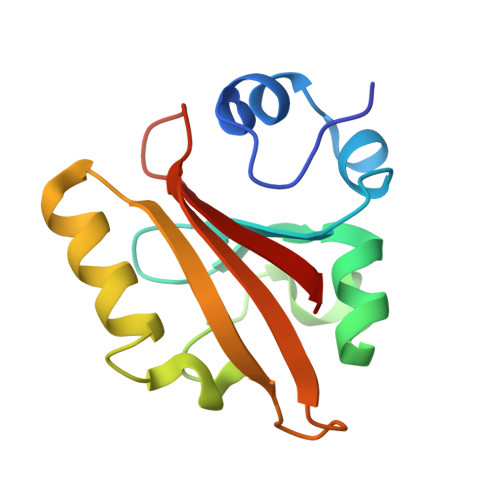
    Compared with batch and vapor diffusion methods, counter diffusion can generate larger and higher-quality protein crystals yielding improved diffraction data and higher-resolution structures. Typically, counter-diffusion experiments are conducted in elongated chambers, such as glass capillaries, and the crystals are either directly measured in the capillary or extracted and mounted at the X-ray beamline. Despite the advantages of counter-diffusion protein crystallization, there are few fixed-target devices that utilize counter diffusion for crystallization. In this article, different designs of user-friendly counter-diffusion chambers are presented which can be used to grow large protein crystals in a 2D polymer microfluidic fixed-target chip. Methods for rapid chip fabrication using commercially available thin-film materials such as Mylar, propyl-ene and Kapton are also detailed. Rules of thumb are provided to tune the nucleation and crystal growth to meet users' needs while minimizing sample consumption. These designs provide a reliable approach to forming large crystals and maintaining their hydration for weeks and even months. This allows ample time to grow, select and preserve the best crystal batches before X-ray beam time. Importantly, the fixed-target microfluidic chip has a low background scatter and can be directly used at beamlines without any crystal handling, enabling crystal quality to be preserved. The approach is demonstrated with serial diffraction of photoactive yellow protein, yielding 1.32 Å resolution at room temperature. Fabrication of this standard microfluidic chip with commercially available thin films greatly simplifies fabrication and provides enhanced stability under vacuum. These advances will further broaden microfluidic fixed-target utilization by crystallographers.


  • Organizational Affiliation
    • Department of Materials Science and Engineering University of California Davis Davis CA95616 USA.

Macromolecules
Find similar proteins by:  (by identity cutoff)  |  3D Structure
Entity ID: 1
MoleculeChains Sequence LengthOrganismDetailsImage
Photoactive yellow protein125Halorhodospira halophilaMutation(s): 0 
Gene Names: pyp
UniProt
Find proteins for P16113 (Halorhodospira halophila)
Explore P16113 
Go to UniProtKB:  P16113
Entity Groups  
Sequence Clusters30% Identity50% Identity70% Identity90% Identity95% Identity100% Identity
UniProt GroupP16113
Sequence Annotations
Expand
  • Reference Sequence
Small Molecules
Ligands 1 Unique
IDChains Name / Formula / InChI Key2D Diagram3D Interactions
HC4
Query on HC4

Download Ideal Coordinates CCD File 
B [auth A]4'-HYDROXYCINNAMIC ACID
C9 H8 O3
NGSWKAQJJWESNS-ZZXKWVIFSA-N
Experimental Data & Validation

Experimental Data

  • Method: X-RAY DIFFRACTION
  • Resolution: 1.32 Å
  • R-Value Free:  0.179 (Depositor), 0.177 (DCC) 
  • R-Value Work:  0.171 (Depositor), 0.170 (DCC) 
  • R-Value Observed: 0.172 (Depositor) 
Space Group: P 65
Unit Cell:
Length ( Å )Angle ( ˚ )
a = 41.194α = 90
b = 41.194β = 90
c = 117.819γ = 120
Software Package:
Software NamePurpose
PHENIXrefinement
xia2data reduction
xia2data scaling
PHENIXphasing

Structure Validation

View Full Validation Report


Ligand Structure Quality Assessment 


Entry History & Funding Information

Deposition Data


Funding OrganizationLocationGrant Number
National Science Foundation (NSF, United States)United States1231306
National Science Foundation (NSF, United States)United States2153503
National Institutes of Health/National Institute of General Medical Sciences (NIH/NIGMS)United StatesR01GM117342
National Institutes of Health/National Institute Of Allergy and Infectious Diseases (NIH/NIAID)United StatesU19 AI144184
Department of Energy (DOE, United States)United StatesP30GM133894

Revision History  (Full details and data files)

  • Version 1.0: 2025-06-04
    Type: Initial release
  • Version 1.1: 2026-03-04
    Changes: Database references