9CBV | pdb_00009cbv

MicroED structure of the human MP20 protein


Experimental Data Snapshot

  • Method: ELECTRON CRYSTALLOGRAPHY
  • Resolution: 3.50 Å
  • R-Value Free: 
    0.351 (Depositor) 
  • R-Value Work: 
    0.332 (Depositor) 
  • R-Value Observed: 
    0.333 (Depositor) 

Starting Model: in silico
View more details

wwPDB Validation   3D Report Full Report


This is version 1.0 of the entry. See complete history


Literature

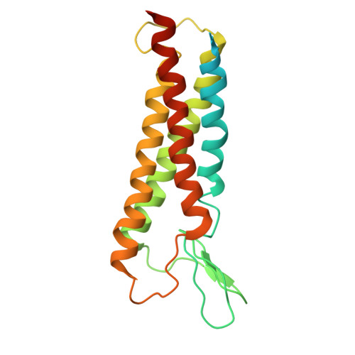
Structure of the lens MP20 mediated adhesive junction.

Nicolas, W.J.Shiriaeva, A.Martynowycz, M.W.Grey, A.C.Ruma, Y.N.Donaldson, P.J.Gonen, T.

(2025) Nat Commun 16: 2977-2977

  • DOI: https://doi.org/10.1038/s41467-025-57903-6
  • Primary Citation of Related Structures:  
    9CBV

  • PubMed Abstract: 

    Human lens fiber membrane intrinsic protein MP20 is the second most abundant membrane protein of the human eye lens. Despite decades of effort its structure and function remained elusive. Here, we determined the MicroED structure of full-length human MP20 in lipidic-cubic phase to a resolution of 3.5 Å. MP20 forms tetramers each of which contain 4 transmembrane α-helices that are packed against one another forming a helical bundle. We find that each MP20 tetramer formed adhesive interactions with an opposing tetramer in a head-to-head fashion. Investigation of MP20 localization in human lenses indicate that in young fiber cells MP20 is initially localized to the cytoplasm in differentiating fiber cells but upon fiber cell maturation is inserted into the plasma membrane, correlating with the restriction of the diffusion of extracellular tracers into the lens. Together these results suggest that MP20 forms lens thin junctions in vivo, confirming its role as a structural protein in the human eye lens essential for its optical transparency.


  • Organizational Affiliation
    • Department of Biological Chemistry, David Geffen School of Medicine, University of California, Los Angeles, CA, USA.

Macromolecules
Find similar proteins by:  (by identity cutoff)  |  3D Structure
Entity ID: 1
MoleculeChains Sequence LengthOrganismDetailsImage
Lens fiber membrane intrinsic protein218Homo sapiensMutation(s): 0 
Gene Names: LIM2
Membrane Entity: Yes 
UniProt & NIH Common Fund Data Resources
Find proteins for P55344 (Homo sapiens)
Explore P55344 
Go to UniProtKB:  P55344
PHAROS:  P55344
GTEx:  ENSG00000105370 
Entity Groups  
Sequence Clusters30% Identity50% Identity70% Identity90% Identity95% Identity100% Identity
UniProt GroupP55344
Sequence Annotations
Expand
  • Reference Sequence
Experimental Data & Validation

Experimental Data

  • Method: ELECTRON CRYSTALLOGRAPHY
  • Resolution: 3.50 Å
  • R-Value Free:  0.351 (Depositor) 
  • R-Value Work:  0.332 (Depositor) 
  • R-Value Observed: 0.333 (Depositor) 
Space Group: P 4 21 2
Unit Cell:
Length ( Å )Angle ( ˚ )
a = 56.18α = 90
b = 56.18β = 90
c = 142.5γ = 90
Software Package:
Software NamePurpose
XSCALEdata scaling
EM Software:
TaskSoftware PackageVersion
MODEL REFINEMENTPHENIX
RECONSTRUCTIONPHENIX

Structure Validation

View Full Validation Report


Entry History & Funding Information

Deposition Data


Funding OrganizationLocationGrant Number
National Institutes of Health/National Eye Institute (NIH/NEI)United StatesP41GM136508
Howard Hughes Medical Institute (HHMI)United States--

Revision History  (Full details and data files)

  • Version 1.0: 2025-05-21
    Type: Initial release