9CAP | pdb_00009cap

Structure of the LPD-3 complex


Experimental Data Snapshot

  • Method: ELECTRON MICROSCOPY
  • Resolution: 2.70 Å
  • Aggregation State: PARTICLE 
  • Reconstruction Method: SINGLE PARTICLE 

wwPDB Validation   3D Report Full Report


This is version 2.0 of the entry. See complete history


Literature

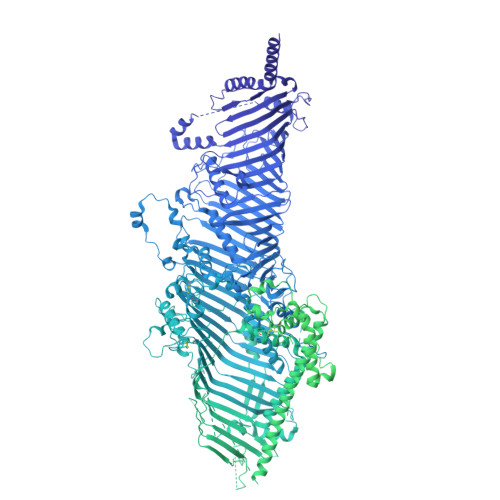
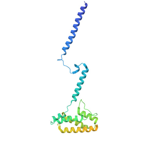
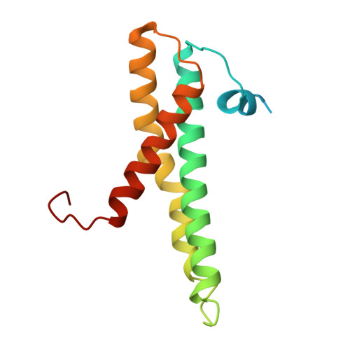
Structural basis of lipid transfer by a bridge-like lipid-transfer protein.

Kang, Y.Lehmann, K.S.Long, H.Jefferson, A.Purice, M.Freeman, M.Clark, S.

(2025) Nature 642: 242-249

  • DOI: https://doi.org/10.1038/s41586-025-08918-y
  • Primary Citation of Related Structures:  
    9CAP

  • PubMed Abstract: 

    Bridge-like lipid-transport proteins (BLTPs) are an evolutionarily conserved family of proteins that localize to membrane-contact sites and are thought to mediate the bulk transfer of lipids from a donor membrane, typically the endoplasmic reticulum, to an acceptor membrane, such as that of the cell or an organelle 1 . Although BLTPs are fundamentally important for a wide array of cellular functions, their architecture, composition and lipid-transfer mechanisms remain poorly characterized. Here we present the subunit composition and the cryogenic electron microscopy structure of the native LPD-3 BLTP complex isolated from transgenic Caenorhabditis elegans. LPD-3 folds into an elongated, rod-shaped tunnel of which the interior is filled with ordered lipid molecules that are coordinated by a track of ionizable residues that line one side of the tunnel. LPD-3 forms a complex with two previously uncharacterized proteins, one of which we have named Spigot and the other of which remains unnamed. Spigot interacts with the N-terminal end of LPD-3 where lipids are expected to enter the tunnel, and experiments in multiple model systems indicate that Spigot has a conserved role in BLTP function. Our LPD-3 complex structural data reveal protein-lipid interactions that suggest a model for how the native LPD-3 complex mediates bulk lipid transport and provides a foundation for mechanistic studies of BLTPs.


  • Organizational Affiliation
    • Vollum Institute, Oregon Health & Science University, Portland, OR, USA.

Macromolecules
Find similar proteins by:  (by identity cutoff)  |  3D Structure
Entity ID: 1
MoleculeChains Sequence LengthOrganismDetailsImage
Bridge-like lipid transfer protein family member 1 C-terminal domain-containing protein4,299Caenorhabditis elegansMutation(s): 0 
UniProt
Find proteins for A0A0K3AWP8 (Caenorhabditis elegans)
Explore A0A0K3AWP8 
Go to UniProtKB:  A0A0K3AWP8
Entity Groups  
Sequence Clusters30% Identity50% Identity70% Identity90% Identity95% Identity100% Identity
UniProt GroupA0A0K3AWP8
Sequence Annotations
Expand
  • Reference Sequence
Find similar proteins by:  (by identity cutoff)  |  3D Structure
Entity ID: 2
MoleculeChains Sequence LengthOrganismDetailsImage
Defect at low temperature protein 1276Caenorhabditis elegansMutation(s): 0 
UniProt
Find proteins for Q4W5H0 (Caenorhabditis elegans)
Explore Q4W5H0 
Go to UniProtKB:  Q4W5H0
Entity Groups  
Sequence Clusters30% Identity50% Identity70% Identity90% Identity95% Identity100% Identity
UniProt GroupQ4W5H0
Sequence Annotations
Expand
  • Reference Sequence
Find similar proteins by:  (by identity cutoff)  |  3D Structure
Entity ID: 3
MoleculeChains Sequence LengthOrganismDetailsImage
Vesicle transport protein143Caenorhabditis elegansMutation(s): 0 
UniProt
Find proteins for Q95QG3 (Caenorhabditis elegans)
Explore Q95QG3 
Go to UniProtKB:  Q95QG3
Entity Groups  
Sequence Clusters30% Identity50% Identity70% Identity90% Identity95% Identity100% Identity
UniProt GroupQ95QG3
Sequence Annotations
Expand
  • Reference Sequence
Small Molecules
Ligands 2 Unique
IDChains Name / Formula / InChI Key2D Diagram3D Interactions
PEE
Query on PEE

Download Ideal Coordinates CCD File 
AA [auth A]
CA [auth A]
D [auth A]
DA [auth A]
E [auth A]
AA [auth A],
CA [auth A],
D [auth A],
DA [auth A],
E [auth A],
EA [auth C],
F [auth A],
G [auth A],
H [auth A],
M [auth A],
N [auth A],
O [auth A],
P [auth A],
U [auth A],
V [auth A],
W [auth A],
X [auth A],
Y [auth A]
1,2-dioleoyl-sn-glycero-3-phosphoethanolamine
C41 H78 N O8 P
MWRBNPKJOOWZPW-NYVOMTAGSA-N
R16
Query on R16

Download Ideal Coordinates CCD File 
BA [auth A]
FA [auth C]
GA [auth C]
I [auth A]
J [auth A]
BA [auth A],
FA [auth C],
GA [auth C],
I [auth A],
J [auth A],
K [auth A],
L [auth A],
Q [auth A],
R [auth A],
S [auth A],
T [auth A],
Z [auth A]
HEXADECANE
C16 H34
DCAYPVUWAIABOU-UHFFFAOYSA-N
Experimental Data & Validation

Experimental Data

  • Method: ELECTRON MICROSCOPY
  • Resolution: 2.70 Å
  • Aggregation State: PARTICLE 
  • Reconstruction Method: SINGLE PARTICLE 
EM Software:
TaskSoftware PackageVersion
MODEL REFINEMENTPHENIX1.21.2_5419
RECONSTRUCTIONcryoSPARC

Structure Validation

View Full Validation Report


Entry History & Funding Information

Deposition Data


Funding OrganizationLocationGrant Number
Howard Hughes Medical Institute (HHMI)United States--

Revision History  (Full details and data files)

  • Version 1.0: 2025-04-23
    Type: Initial release
  • Version 1.1: 2025-05-07
    Changes: Data collection, Database references
  • Version 1.2: 2025-06-18
    Changes: Data collection, Database references
  • Version 2.0: 2025-12-10
    Type: Coordinate replacement
    Reason: Model completeness
    Changes: Advisory, Atomic model, Data collection, Database references, Derived calculations, Polymer sequence, Source and taxonomy, Structure summary