9C6S | pdb_00009c6s

18-mer blood cell-specific tubulin in complex with Cryptophycin-52

  • Classification: CELL CYCLE
  • Organism(s): Gallus gallus
  • Mutation(s): No 

  • Deposited: 2024-06-08 Released: 2025-01-01 
  • Deposition Author(s): Montecinos, F.
  • Funding Organization(s): National Institutes of Health/National Institute of Arthritis and Musculoskeletal and Skin Diseases (NIH/NIAMS)

Experimental Data Snapshot

  • Method: ELECTRON MICROSCOPY
  • Resolution: 3.52 Å
  • Aggregation State: PARTICLE 
  • Reconstruction Method: SINGLE PARTICLE 

wwPDB Validation 3D Report Full Report

Validation slider image for 9C6S

This is version 1.3 of the entry. See complete history

Literature

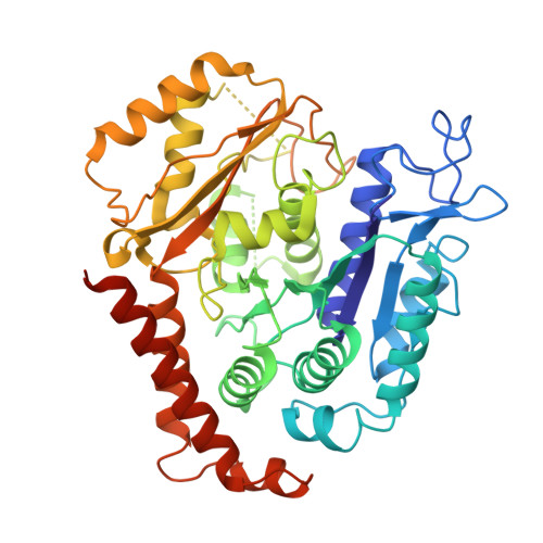
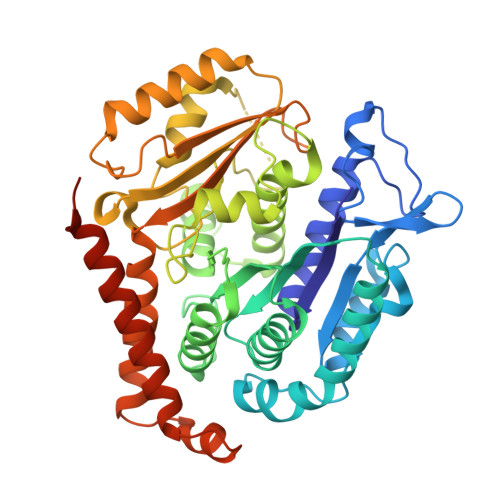
Structure of blood cell-specific tubulin and demonstration of dimer spacing compaction in a single protofilament.

Montecinos, F.Eren, E.Watts, N.R.Sackett, D.L.Wingfield, P.T.

(2024) J Biological Chem 301: 108132-108132

  • DOI: https://doi.org/10.1016/j.jbc.2024.108132
  • Primary Citation Related Structures: 
    9C6R, 9C6S

  • PubMed Abstract: 

    Microtubule (MT) function plasticity originates from its composition of α- and β-tubulin isotypes and the post-translational modifications of both subunits. Aspects such as MT assembly dynamics, structure, and anticancer drug binding can be modulated by αβ-tubulin heterogeneity. However, the exact molecular mechanism regulating these aspects is only partially understood. A recent insight is the discovery of expansion and compaction of the MT lattice, which can occur via fine modulation of dimer longitudinal spacing mediated by GTP hydrolysis, taxol binding, protein binding, or isotype composition. Here, we report the first structure of the blood cell-specific α1/β1-tubulin isolated from the marginal band of chicken erythrocytes (ChET) determined to a resolution of 3.2 Å by cryo-EM. We show that ChET rings induced with Cryptophycin-52 (Cp-52) are smaller in diameter than HeLa tubulin (HeLaT) rings induced with Cp-52 and composed of the same number of heterodimers. We observe compacted interdimer and intradimer curved protofilament interfaces, characterized by shorter distances between ChET subunits and accompanied by conformational changes in the β-tubulin subunit. The compacted ChET interdimer interface brings more residues near the Cp-52 binding site. We measured the Cp-52 apparent binding affinities of ChET and HeLaT by mass photometry, observing small differences, and detected the intermediates of the ring assembly reaction. These findings demonstrate that compaction/expansion of dimer spacing can occur in a single protofilament context and that the subtle structural differences between tubulin isotypes can modulate tubulin small molecule binding.


  • Organizational Affiliation
    • Protein Expression Laboratory, National Institute of Arthritis and Musculoskeletal and Skin Diseases, National Institutes of Health, Bethesda, Maryland, USA.

Macromolecule Content 

  • Total Structure Weight: 918.23 kDa 
  • Atom Count: 61,137 
  • Modeled Residue Count: 7,677 
  • Deposited Residue Count: 8,073 
  • Unique protein chains: 2

Macromolecules

Find similar proteins by:|  3D Structure
Entity ID: 1
MoleculeChains  Sequence LengthOrganismDetailsImage
Detyrosinated tubulin alpha-1A chain451Gallus gallusMutation(s): 0 
EC: 3.6.5
UniProt
Find proteins for P02552 (Gallus gallus)
Explore P02552 
Go to UniProtKB:  P02552
Entity Groups
Sequence Clusters30% Identity50% Identity70% Identity90% Identity95% Identity100% Identity
UniProt GroupP02552
Sequence Annotations
Expand
Reference Sequence
Find similar proteins by:|  3D Structure
Entity ID: 2
MoleculeChains  Sequence LengthOrganismDetailsImage
Tubulin beta-6 chain446Gallus gallusMutation(s): 0 
UniProt
Find proteins for P09207 (Gallus gallus)
Explore P09207 
Go to UniProtKB:  P09207
Entity Groups
Sequence Clusters30% Identity50% Identity70% Identity90% Identity95% Identity100% Identity
UniProt GroupP09207
Sequence Annotations
Expand
Reference Sequence

Small Molecules

Ligands 3 Unique
IDChains Name / Formula / InChI Key2D Diagram3D Interactions
YGY
(Subject of Investigation/LOI)

Query on YGY


Download:Ideal Coordinates CCD File
AA [auth J]
DA [auth N]
GA [auth R]
JA [auth V]
MA [auth Z]
AA [auth J],
DA [auth N],
GA [auth R],
JA [auth V],
MA [auth Z],
PA [auth d],
SA [auth h],
U [auth B],
X [auth F]
Cryptophycin 52
C36 H45 Cl N2 O8
LSXOBYNBRKOTIQ-HZMFLOLBSA-N
GTP
(Subject of Investigation/LOI)

Query on GTP


Download:Ideal Coordinates CCD File
BA [auth M]
EA [auth Q]
HA [auth U]
KA [auth Y]
NA [auth c]
BA [auth M],
EA [auth Q],
HA [auth U],
KA [auth Y],
NA [auth c],
QA [auth g],
S [auth A],
V [auth C],
Y [auth I]
GUANOSINE-5'-TRIPHOSPHATE
C10 H16 N5 O14 P3
XKMLYUALXHKNFT-UUOKFMHZSA-N
GDP
(Subject of Investigation/LOI)

Query on GDP


Download:Ideal Coordinates CCD File
CA [auth N]
FA [auth R]
IA [auth V]
LA [auth Z]
OA [auth d]
CA [auth N],
FA [auth R],
IA [auth V],
LA [auth Z],
OA [auth d],
RA [auth h],
T [auth B],
W [auth F],
Z [auth J]
GUANOSINE-5'-DIPHOSPHATE
C10 H15 N5 O11 P2
QGWNDRXFNXRZMB-UUOKFMHZSA-N

Experimental Data & Validation

Experimental Data

  • Method: ELECTRON MICROSCOPY
  • Resolution: 3.52 Å
  • Aggregation State: PARTICLE 
  • Reconstruction Method: SINGLE PARTICLE 
EM Software:
TaskSoftware PackageVersion
MODEL REFINEMENTPHENIX

Structure Validation

View Full Validation Report


Entry History 

& Funding Information

Deposition Data


Funding OrganizationLocationGrant Number
National Institutes of Health/National Institute of Arthritis and Musculoskeletal and Skin Diseases (NIH/NIAMS)United StatesIntramural Research Program

Revision History  (Full details and data files)

  • Version 1.0: 2025-01-01
    Type: Initial release
  • Version 1.1: 2025-01-08
    Changes: Data collection, Database references
  • Version 1.2: 2025-01-29
    Changes: Data collection, Database references
  • Version 1.3: 2025-05-14
    Changes: Data collection