9C5G | pdb_00009c5g

Structure of R. leguminosarum CapW bound to single-stranded DNA


Experimental Data Snapshot

  • Method: X-RAY DIFFRACTION
  • Resolution: 2.77 Å
  • R-Value Free: 
    0.279 (Depositor), 0.278 (DCC) 
  • R-Value Work: 
    0.230 (Depositor), 0.230 (DCC) 
  • R-Value Observed: 
    0.232 (Depositor) 

Starting Model: in silico
View more details

wwPDB Validation 3D Report Full Report

Validation slider image for 9C5G

This is version 1.2 of the entry. See complete history

Literature

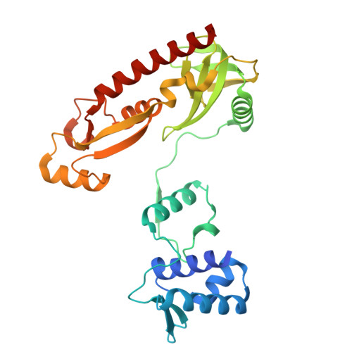
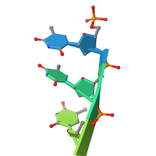
Bacterial WYL domain transcriptional repressors sense single-stranded DNA to control gene expression.

Blankenchip, C.L.Corbett, K.D.

(2024) Nucleic Acids Res 52: 13723-13732

  • DOI: https://doi.org/10.1093/nar/gkae1101
  • Primary Citation Related Structures: 
    9C5G

  • PubMed Abstract: 

    Bacteria encode a wide array of immune systems to protect themselves against ubiquitous bacteriophages and foreign DNA elements. While these systems' molecular mechanisms are becoming increasingly well known, their regulation remains poorly understood. Here, we show that an immune system-associated transcriptional repressor of the wHTH-WYL-WCX family, CapW, directly binds single-stranded DNA to sense DNA damage and activate expression of its associated immune system. We show that CapW mediates increased expression of a reporter gene in response to DNA damage in a host cell. CapW directly binds single-stranded DNA by-products of DNA repair through its WYL domain, causing a conformational change that releases the protein from double-stranded DNA. In an Escherichia coli CBASS system with an integrated capW gene, we find that CapW-mediated transcriptional activation is important for this system's ability to prevent induction of a λ prophage. Overall, our data reveal the molecular mechanisms of WYL-domain transcriptional repressors, and provide an example of how bacteria can balance the protective benefits of carrying anti-phage immune systems against the inherent risk of these systems' aberrant activation.


  • Organizational Affiliation
    • Biomedical Sciences Graduate Program, University of California, San Diego, 9500 Gilman Dr. La Jolla, CA 92093, USA.

Macromolecule Content 

  • Total Structure Weight: 74.56 kDa 
  • Atom Count: 4,880 
  • Modeled Residue Count: 584 
  • Deposited Residue Count: 612 
  • Unique protein chains: 1
  • Unique nucleic acid chains: 1

Macromolecules


Find similar proteins by:|  3D Structure
Entity ID: 1
MoleculeChains  Sequence LengthOrganismDetailsImage
CapW
A, B
299Rhizobium leguminosarumMutation(s): 0 
Find similar nucleic acids by:  Sequence
Entity ID: 2
MoleculeChains LengthOrganismImage
DNA (5'-D(P*TP*TP*T)-3')
C, D
7Escherichia coli
Sequence Annotations
Expand
Reference Sequence

Small Molecules

Ligands 1 Unique
IDChains Name / Formula / InChI Key2D Diagram3D Interactions
SO4

Query on SO4


Download:Ideal Coordinates CCD File
E [auth B]SULFATE ION
O4 S
QAOWNCQODCNURD-UHFFFAOYSA-L

Experimental Data & Validation

Experimental Data

  • Method: X-RAY DIFFRACTION
  • Resolution: 2.77 Å
  • R-Value Free:  0.279 (Depositor), 0.278 (DCC) 
  • R-Value Work:  0.230 (Depositor), 0.230 (DCC) 
  • R-Value Observed: 0.232 (Depositor) 
Space Group: P 1 21 1
Unit Cell:
Length ( Å )Angle ( ˚ )
a = 77.955α = 90
b = 62.008β = 93.481
c = 82.335γ = 90
Software Package:
Software NamePurpose
PHENIXrefinement
XDSdata reduction
Aimlessdata scaling
PHASERphasing

Structure Validation

View Full Validation Report


Entry History 

& Funding Information

Deposition Data


Funding OrganizationLocationGrant Number
National Institutes of Health/National Institute of General Medical Sciences (NIH/NIGMS)United StatesR35 GM144121

Revision History  (Full details and data files)

  • Version 1.0: 2024-10-30
    Type: Initial release
  • Version 1.1: 2024-12-11
    Changes: Database references
  • Version 1.2: 2025-01-01
    Changes: Database references