9C2F | pdb_00009c2f

Structure of Calcium-Sensing Receptor in complex with positive allosteric modulator '54149


Experimental Data Snapshot

  • Method: ELECTRON MICROSCOPY
  • Resolution: 2.80 Å
  • Aggregation State: PARTICLE 
  • Reconstruction Method: SINGLE PARTICLE 

wwPDB Validation 3D Report Full Report

Validation slider image for 9C2F

This is version 1.2 of the entry. See complete history

Literature

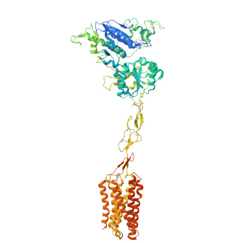
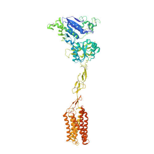
Large library docking identifies positive allosteric modulators of the calcium-sensing receptor.

Liu, F.Wu, C.G.Tu, C.L.Glenn, I.Meyerowitz, J.Kaplan, A.L.Lyu, J.Cheng, Z.Tarkhanova, O.O.Moroz, Y.S.Irwin, J.J.Chang, W.Shoichet, B.K.Skiniotis, G.

(2024) Science 385: eado1868-eado1868

  • DOI: https://doi.org/10.1126/science.ado1868
  • Primary Citation Related Structures: 
    9C1P, 9C2F

  • PubMed Abstract: 

    Positive allosteric modulator (PAM) drugs enhance the activation of the calcium-sensing receptor (CaSR) and suppress parathyroid hormone (PTH) secretion. Unfortunately, these hyperparathyroidism-treating drugs can induce hypocalcemia and arrhythmias. Seeking improved modulators, we docked libraries of 2.7 million and 1.2 billion molecules against the CaSR structure. The billion-molecule docking found PAMs with a 2.7-fold higher hit rate than the million-molecule library, with hits up to 37-fold more potent. Structure-based optimization led to nanomolar leads. In ex vivo organ assays, one of these PAMs was 100-fold more potent than the standard of care, cinacalcet, and reduced serum PTH levels in mice without the hypocalcemia typical of CaSR drugs. As determined from cryo-electron microscopy structures, the PAMs identified here promote CaSR conformations that more closely resemble the activated state than those induced by the established drugs.


  • Organizational Affiliation
    • Department of Pharmaceutical Chemistry, University of California, San Francisco, San Francisco, CA 94143, USA.

Macromolecule Content 

  • Total Structure Weight: 217.06 kDa 
  • Atom Count: 12,163 
  • Modeled Residue Count: 1,540 
  • Deposited Residue Count: 1,898 
  • Unique protein chains: 2

Macromolecules

Find similar proteins by:|  3D Structure
Entity ID: 1
MoleculeChains  Sequence LengthOrganismDetailsImage
Extracellular calcium-sensing receptor959Homo sapiensMutation(s): 0 
Gene Names: CASRGPRC2APCAR1
Membrane Entity: Yes 
UniProt & NIH Common Fund Data Resources
Find proteins for P41180 (Homo sapiens)
Explore P41180 
Go to UniProtKB:  P41180
PHAROS:  P41180
GTEx:  ENSG00000036828 
Entity Groups
Sequence Clusters30% Identity50% Identity70% Identity90% Identity95% Identity100% Identity
UniProt GroupP41180
Glycosylation
Glycosylation Sites: 3Go to GlyGen: P41180-1
Sequence Annotations
Expand
Reference Sequence
Find similar proteins by:|  3D Structure
Entity ID: 2
MoleculeChains  Sequence LengthOrganismDetailsImage
Extracellular calcium-sensing receptor939Homo sapiensMutation(s): 0 
Gene Names: CASRGPRC2APCAR1
Membrane Entity: Yes 
UniProt & NIH Common Fund Data Resources
Find proteins for P41180 (Homo sapiens)
Explore P41180 
Go to UniProtKB:  P41180
PHAROS:  P41180
GTEx:  ENSG00000036828 
Entity Groups
Sequence Clusters30% Identity50% Identity70% Identity90% Identity95% Identity100% Identity
UniProt GroupP41180
Glycosylation
Glycosylation Sites: 3Go to GlyGen: P41180-1
Sequence Annotations
Expand
Reference Sequence

Oligosaccharides

Help  
Entity ID: 3
MoleculeChains Length2D Diagram GlycosylationD Interactions
2-acetamido-2-deoxy-beta-D-glucopyranose-(1-4)-2-acetamido-2-deoxy-beta-D-glucopyranoseC [auth H]2N-Glycosylation
Glycosylation Resources
GlyTouCan: G42666HT
GlyCosmos: G42666HT
GlyGen: G42666HT

Small Molecules

Ligands 5 Unique
IDChains Name / Formula / InChI Key2D Diagram3D Interactions
A1ATX
(Subject of Investigation/LOI)

Query on A1ATX


Download:Ideal Coordinates CCD File
J [auth A],
V [auth B]
(1R)-1-(2H-1,3-benzodioxol-4-yl)-N-[2-(1,2-benzothiazol-3-yl)ethyl]ethan-1-amine
C18 H18 N2 O2 S
NSLXTKXZHISXFJ-GFCCVEGCSA-N
NAG

Query on NAG


Download:Ideal Coordinates CCD File
D [auth A],
K [auth A],
L [auth A],
N [auth B],
O [auth B]
2-acetamido-2-deoxy-beta-D-glucopyranose
C8 H15 N O6
OVRNDRQMDRJTHS-FMDGEEDCSA-N
TRP

Query on TRP


Download:Ideal Coordinates CCD File
H [auth A],
P [auth B]
TRYPTOPHAN
C11 H12 N2 O2
QIVBCDIJIAJPQS-VIFPVBQESA-N
PO4

Query on PO4


Download:Ideal Coordinates CCD File
I [auth A],
U [auth B]
PHOSPHATE ION
O4 P
NBIIXXVUZAFLBC-UHFFFAOYSA-K
CA

Query on CA


Download:Ideal Coordinates CCD File
E [auth A]
F [auth A]
G [auth A]
M [auth B]
Q [auth B]
E [auth A],
F [auth A],
G [auth A],
M [auth B],
Q [auth B],
R [auth B],
S [auth B],
T [auth B]
CALCIUM ION
Ca
BHPQYMZQTOCNFJ-UHFFFAOYSA-N

Experimental Data & Validation

Experimental Data

  • Method: ELECTRON MICROSCOPY
  • Resolution: 2.80 Å
  • Aggregation State: PARTICLE 
  • Reconstruction Method: SINGLE PARTICLE 
EM Software:
TaskSoftware PackageVersion
MODEL REFINEMENTPHENIX

Structure Validation

View Full Validation Report


Entry History 

& Funding Information

Deposition Data


Funding OrganizationLocationGrant Number
National Institutes of Health/National Institute of Neurological Disorders and Stroke (NIH/NINDS)United StatesRO1 NS122394
National Institutes of Health/National Institute of Neurological Disorders and Stroke (NIH/NINDS)United StatesRO1 DK132902

Revision History  (Full details and data files)

  • Version 1.0: 2024-10-02
    Type: Initial release
  • Version 1.1: 2024-11-06
    Changes: Data collection, Structure summary
  • Version 1.2: 2025-05-28
    Changes: Data collection