


In silico Self-assembling Protein Nanoparticles Optimization for Protein Antigen display
Cappelli, L., Wahome, N., Cinelli, P., Giusti, F., Seraj, N., Cartocci, E., Harshbarger, W., Delany, I., Cozzi, R.To be published.
Experimental Data Snapshot
Starting Model: experimental
View more details
wwPDB Validation 3D Report Full Report

Entity ID: 1 | |||||
|---|---|---|---|---|---|
| Molecule | Chains | Sequence Length | Organism | Details | Image |
| Flavoprotein | 79 | Mycobacterium tuberculosis | Mutation(s): 0 Gene Names: Rv1498A | 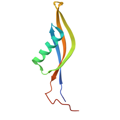 | |
UniProt | |||||
Entity Groups | |||||
| Sequence Clusters | 30% Identity50% Identity70% Identity90% Identity95% Identity100% Identity | ||||
| UniProt Group | I6XY36 | ||||
Sequence AnnotationsExpand | |||||
Reference Sequence | |||||
| Length ( Å ) | Angle ( ˚ ) |
|---|---|
| a = 139.183 | α = 90 |
| b = 139.183 | β = 90 |
| c = 139.183 | γ = 90 |
| Software Name | Purpose |
|---|---|
| PHENIX | refinement |
| Coot | model building |
| XDS | data reduction |
| PHENIX | phasing |
| HKL-2000 | data scaling |
| Funding Organization | Location | Grant Number |
|---|---|---|
| Other private | United States | -- |


