9BOP | pdb_00009bop

A broadly-neutralizing antibody against Ebolavirus glycoprotein that can potentiate the breadth and neutralization potency of other anti-glycoprotein antibodies


Experimental Data Snapshot

  • Method: ELECTRON MICROSCOPY
  • Resolution: 3.00 Å
  • Aggregation State: PARTICLE 
  • Reconstruction Method: SINGLE PARTICLE 

wwPDB Validation 3D Report Full Report

Validation slider image for 9BOP

This is version 1.1 of the entry. See complete history

Literature

A broadly-neutralizing antibody against Orthoebolavirus glycoprotein that potentiates the breadth and neutralization of other antibodies.

Donnellan, F.R.Rayaprolu, V.Rijal, P.O'Dowd, V.Parvate, A.Callaway, H.Hariharan, C.Parekh, D.Hui, S.Shaffer, K.C.L.Avalos, R.D.Hastie, K.M.Schimanski, L.Muller-Krauter, H.Strecker, T.Balaram, A.Halfmann, P.Saphire, E.O.Lightwood, D.J.Townsend, A.R.Draper, S.J.

(2026) Npj Viruses 

  • DOI: https://doi.org/10.1038/s44298-026-00192-7
  • Primary Citation Related Structures: 
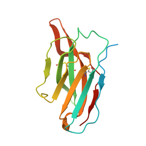
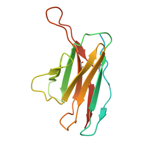
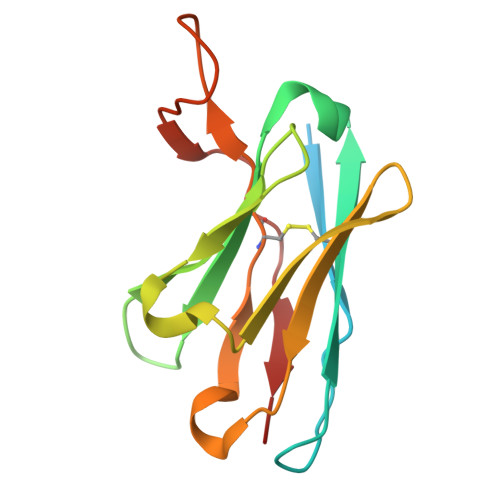
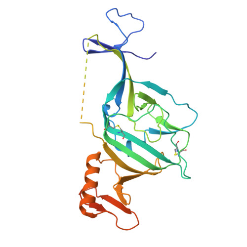
    9BOP

  • PubMed Abstract: 

    Ebolavirus disease (EVD) is caused by multiple species of orthoebolavirus. Monoclonal antibodies (mAbs) against the virus glycoprotein (GP) are the only class of therapeutic approved for treatment of EVD caused by Orthoebolavirus zairense (Ebola virus, EBOV). Therefore, mAbs targeting multiple orthoebolavirus species may represent the next generation of EVD therapeutics. Broadly reactive anti-GP mAbs were produced; among these, mAbs 11886 and 11883 were broadly neutralizing in vitro. A 3.0 Å cryo-electron microscopy structure of EBOV GP bound to both mAbs shows that 11886 binds a novel epitope bridging the glycan cap (GC), 3 10 pocket and GP2 N-terminus, whereas 11883 binds the receptor binding region (RBR) and GC. In vitro, 11886 synergized with a range of mAbs with epitope specificities spanning the RBR/GC, including 11883. Notably, 11886 increased the breadth of neutralization by partner mAbs against different orthoebolavirus species. These data provide a strategic route to design improved mAb-based next-generation EVD therapeutics.


  • Organizational Affiliation
    • Department of Biochemistry, Dorothy Crowfoot Hodgkin Building, University of Oxford, Oxford, UK.

Macromolecule Content 

  • Total Structure Weight: 298.51 kDa 
  • Atom Count: 16,858 
  • Modeled Residue Count: 2,162 
  • Deposited Residue Count: 2,680 
  • Unique protein chains: 6

Macromolecules

Find similar proteins by:|  3D Structure
Entity ID: 1
MoleculeChains  Sequence LengthOrganismDetailsImage
11883 heavy chainA,
I [auth C],
J [auth E]
148Oryctolagus cuniculusMutation(s): 0 
Find similar proteins by:|  3D Structure
Entity ID: 2
MoleculeChains  Sequence LengthOrganismDetailsImage
11883 light chainB,
K [auth D],
L [auth F]
140Oryctolagus cuniculusMutation(s): 0 
Find similar proteins by:|  3D Structure
Entity ID: 3
MoleculeChains  Sequence LengthOrganismDetailsImage
11886 heavy chainC [auth M],
E [auth O]
146Oryctolagus cuniculusMutation(s): 0 
Find similar proteins by:|  3D Structure
Entity ID: 4
MoleculeChains  Sequence LengthOrganismDetailsImage
11886 light chainD [auth N],
F [auth P]
141Oryctolagus cuniculusMutation(s): 0 
Find similar proteins by:|  3D Structure
Entity ID: 5
MoleculeChains  Sequence LengthOrganismDetailsImage
Virion spike glycoprotein GP1G [auth S],
O [auth T],
P [auth U]
279OrthoebolavirusMutation(s): 0 
Gene Names: GP
UniProt
Find proteins for A0A8G0KJC2 (Zaire ebolavirus)
Explore A0A8G0KJC2 
Go to UniProtKB:  A0A8G0KJC2
Entity Groups
Sequence Clusters30% Identity50% Identity70% Identity90% Identity95% Identity100% Identity
UniProt GroupA0A8G0KJC2
Glycosylation
Glycosylation Sites: 2
Sequence Annotations
Expand
Reference Sequence
Find similar proteins by:|  3D Structure
Entity ID: 6
MoleculeChains  Sequence LengthOrganismDetailsImage
Virion spike glycoprotein GP2H [auth V],
M [auth W],
N [auth X]
135OrthoebolavirusMutation(s): 0 
Gene Names: GP
UniProt
Find proteins for A0A8G0KMX8 (Zaire ebolavirus)
Explore A0A8G0KMX8 
Go to UniProtKB:  A0A8G0KMX8
Entity Groups
Sequence Clusters30% Identity50% Identity70% Identity90% Identity95% Identity100% Identity
UniProt GroupA0A8G0KMX8
Glycosylation
Glycosylation Sites: 1
Sequence Annotations
Expand
Reference Sequence

Oligosaccharides

Help  
Entity ID: 7
MoleculeChains Length2D Diagram GlycosylationD Interactions
alpha-D-mannopyranose-(1-3)-[alpha-D-mannopyranose-(1-6)]beta-D-mannopyranose-(1-4)-2-acetamido-2-deoxy-beta-D-glucopyranose-(1-4)-2-acetamido-2-deoxy-beta-D-glucopyranoseQ [auth G],
R [auth H],
S [auth I]
5N-Glycosylation
Glycosylation Resources
GlyTouCan: G22768VO
GlyCosmos: G22768VO
GlyGen: G22768VO

Small Molecules

Ligands 1 Unique
IDChains Name / Formula / InChI Key2D Diagram3D Interactions
NAG
(Subject of Investigation/LOI)

Query on NAG


Download:Ideal Coordinates CCD File
AA [auth U]
BA [auth U]
T [auth S]
U [auth S]
V [auth S]
AA [auth U],
BA [auth U],
T [auth S],
U [auth S],
V [auth S],
W [auth T],
X [auth T],
Y [auth T],
Z [auth U]
2-acetamido-2-deoxy-beta-D-glucopyranose
C8 H15 N O6
OVRNDRQMDRJTHS-FMDGEEDCSA-N

Experimental Data & Validation

Experimental Data

  • Method: ELECTRON MICROSCOPY
  • Resolution: 3.00 Å
  • Aggregation State: PARTICLE 
  • Reconstruction Method: SINGLE PARTICLE 
EM Software:
TaskSoftware PackageVersion
RECONSTRUCTIONcryoSPARC
MODEL REFINEMENTPHENIX
MODEL REFINEMENTCoot

Structure Validation

View Full Validation Report


Entry History 

& Funding Information

Deposition Data


Funding OrganizationLocationGrant Number
Medical Research Council (MRC, United Kingdom)United KingdomMR/N01796X/1

Revision History  (Full details and data files)

  • Version 1.0: 2025-11-05
    Type: Initial release
  • Version 1.1: 2026-05-20
    Changes: Data collection, Database references, Structure summary