9BOI | pdb_00009boi

Cryo-EM structure of human Spns1 in complex with LPC (18:1)


Experimental Data Snapshot

  • Method: ELECTRON MICROSCOPY
  • Resolution: 3.22 Å
  • Aggregation State: PARTICLE 
  • Reconstruction Method: SINGLE PARTICLE 

wwPDB Validation   3D Report Full Report


This is version 1.1 of the entry. See complete history


Literature

Molecular basis of Spns1-mediated lysophospholipid transport from the lysosome.

Chen, H.Ha, H.T.T.Elghobashi-Meinhardt, N.Le, N.A.Schmiege, P.Nguyen, L.N.Li, X.

(2025) Proc Natl Acad Sci U S A 122: e2409596121-e2409596121

  • DOI: https://doi.org/10.1073/pnas.2409596121
  • Primary Citation of Related Structures:  
    9BOI

  • PubMed Abstract: 

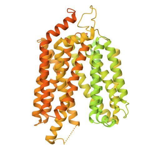
    Spns1 mediates the rate-limiting efflux of lysophospholipids from the lysosome to the cytosol. Deficiency of Spns1 is associated with embryonic senescence, as well as liver and skeletal muscle atrophy in animal models. However, the mechanisms by which Spns1 transports lysophospholipid and proton sensing remain unclear. Here, we present a cryogenic electron microscopy structure of human Spns1 in lysophosphatidylcholine (LPC)-bound lumen-facing conformation. Notably, LPC snugly binds within the luminal-open cavity, where the molecular dynamics simulations reveal that LPC presents a propensity to enter between transmembrane-helices (TM) 5 and 8. Structural comparisons and cell-based transport assays uncover several pivotal residues at TM 5/8 that orchestrate the transport cycle, which are unique to Spns1. Furthermore, we identify a five-residue network that is crucial for proton-sensing by Spns1. Transference of these network residues to Spns2, a sphingosine-1-phosphate uniporter, causes the chimeric Spns2 to be low pH dependent. Our results reveal molecular insights into lysosomal LPC transport and the proton-sensing mechanism by Spns1.


  • Organizational Affiliation
    • Department of Molecular Genetics, University of Texas Southwestern Medical Center, Dallas, TX 75390.

Macromolecules
Find similar proteins by:  (by identity cutoff)  |  3D Structure
Entity ID: 1
MoleculeChains Sequence LengthOrganismDetailsImage
mVenus,Maltose/maltodextrin-binding periplasmic protein,Protein spinster homolog 1,Designed ankyrin repeat protein (DARPin)1,275Aequorea victoriaEscherichia coliHomo sapiensMutation(s): 0 
Gene Names: GFPmalESPNS1SPIN1PP20300
Membrane Entity: Yes 
UniProt & NIH Common Fund Data Resources
Find proteins for P0AEX9 (Escherichia coli (strain K12))
Explore P0AEX9 
Go to UniProtKB:  P0AEX9
Find proteins for P42212 (Aequorea victoria)
Explore P42212 
Go to UniProtKB:  P42212
Find proteins for Q9H2V7 (Homo sapiens)
Explore Q9H2V7 
Go to UniProtKB:  Q9H2V7
PHAROS:  Q9H2V7
GTEx:  ENSG00000169682 
Entity Groups  
Sequence Clusters30% Identity50% Identity70% Identity90% Identity95% Identity100% Identity
UniProt GroupsQ9H2V7P42212P0AEX9
Sequence Annotations
Expand
  • Reference Sequence
Small Molecules
Ligands 1 Unique
IDChains Name / Formula / InChI Key2D Diagram3D Interactions
42H (Subject of Investigation/LOI)
Query on 42H

Download Ideal Coordinates CCD File 
B [auth A](4R,7R,18Z)-4,7-dihydroxy-N,N,N-trimethyl-10-oxo-3,5,9-trioxa-4-phosphaheptacos-18-en-1-aminium 4-oxide
C26 H53 N O7 P
YAMUFBLWGFFICM-PTGWMXDISA-O
Experimental Data & Validation

Experimental Data

  • Method: ELECTRON MICROSCOPY
  • Resolution: 3.22 Å
  • Aggregation State: PARTICLE 
  • Reconstruction Method: SINGLE PARTICLE 
EM Software:
TaskSoftware PackageVersion
MODEL REFINEMENTPHENIX1.16

Structure Validation

View Full Validation Report


Entry History & Funding Information

Deposition Data


Funding OrganizationLocationGrant Number
National Institutes of Health/National Heart, Lung, and Blood Institute (NIH/NHLBI)United StatesP01 HL160487
National Institutes of Health/National Institute of General Medical Sciences (NIH/NIGMS)United StatesR01 GM135343
Welch FoundationUnited StatesI-1957

Revision History  (Full details and data files)

  • Version 1.0: 2025-01-01
    Type: Initial release
  • Version 1.1: 2025-01-22
    Changes: Data collection, Database references