9BIY | pdb_00009biy

Crystal structure of the periplasmic domain of IgaA from Escherichia coli


Experimental Data Snapshot

  • Method: X-RAY DIFFRACTION
  • Resolution: 1.80 Å
  • R-Value Free: 
    0.276 (Depositor), 0.277 (DCC) 
  • R-Value Work: 
    0.240 (Depositor), 0.241 (DCC) 
  • R-Value Observed: 
    0.242 (Depositor) 

wwPDB Validation 3D Report Full Report

Validation slider image for 9BIY

This is version 1.3 of the entry. See complete history

Literature

Molecular insights into the initiation step of the Rcs signaling pathway.

Watanabe, N.Savchenko, A.

(2024) Structure 32: 1381

  • DOI: https://doi.org/10.1016/j.str.2024.06.003
  • Primary Citation Related Structures: 
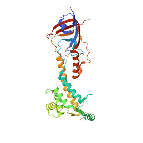
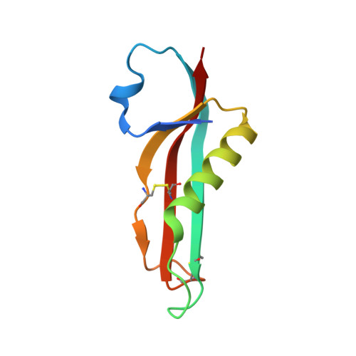
    9BIY, 9BIZ, 9BJ0

  • PubMed Abstract: 

    The Rcs pathway is repressed by the inner membrane protein IgaA under non-stressed conditions. This repression is hypothesized to be relieved by the binding of the outer membrane-anchored RcsF to IgaA. However, the precise mechanism by which RcsF binding triggers the signaling remains unclear. Here, we present the 1.8 Å resolution crystal structure capturing the interaction between IgaA and RcsF. Our comparative structural analysis, examining both the bound and unbound states of the periplasmic domain of IgaA (IgaAp), highlights rotational flexibility within IgaAp. Conversely, the conformation of RcsF remains unchanged upon binding. Our in vivo and in vitro studies do not support the model of a stable complex involving RcsF, IgaAp, and RcsDp. Instead, we demonstrate that the elements beyond IgaAp play a role in the interaction between IgaA and RcsD. These findings collectively allow us to propose a potential mechanism for the signaling across the inner membrane through IgaA.


  • Organizational Affiliation
    • Department of Microbiology, Immunology and Infectious Diseases, University of Calgary, Calgary, AB, Canada; Center for Structural Biology for Infectious Diseases (CSBID) Chicago, IL, USA.

Macromolecule Content 

  • Total Structure Weight: 39.23 kDa 
  • Atom Count: 2,868 
  • Modeled Residue Count: 347 
  • Deposited Residue Count: 363 
  • Unique protein chains: 2

Macromolecules

Find similar proteins by:|  3D Structure
Entity ID: 1
MoleculeChains  Sequence LengthOrganismDetailsImage
Intracellular growth attenuator protein igaA275Escherichia coliMutation(s): 0 
Gene Names: GP975_16005
UniProt
Find proteins for P45800 (Escherichia coli (strain K12))
Explore P45800 
Go to UniProtKB:  P45800
Entity Groups
Sequence Clusters30% Identity50% Identity70% Identity90% Identity95% Identity100% Identity
UniProt GroupP45800
Sequence Annotations
Expand
Reference Sequence
Find similar proteins by:|  3D Structure
Entity ID: 2
MoleculeChains  Sequence LengthOrganismDetailsImage
Outer membrane lipoprotein RcsF88Escherichia coliMutation(s): 0 
Gene Names: rcsFNCTC10865_05029
UniProt
Find proteins for P69411 (Escherichia coli (strain K12))
Explore P69411 
Go to UniProtKB:  P69411
Entity Groups
Sequence Clusters30% Identity50% Identity70% Identity90% Identity95% Identity100% Identity
UniProt GroupP69411
Sequence Annotations
Expand
Reference Sequence

Small Molecules

Modified Residues  1 Unique
IDChains TypeFormula2D DiagramParent
MSE
Query on MSE
A
L-PEPTIDE LINKINGC5 H11 N O2 SeMET

Experimental Data & Validation

Experimental Data

  • Method: X-RAY DIFFRACTION
  • Resolution: 1.80 Å
  • R-Value Free:  0.276 (Depositor), 0.277 (DCC) 
  • R-Value Work:  0.240 (Depositor), 0.241 (DCC) 
  • R-Value Observed: 0.242 (Depositor) 
Space Group: C 1 2 1
Unit Cell:
Length ( Å )Angle ( ˚ )
a = 78.382α = 90
b = 38.266β = 94.965
c = 120.068γ = 90
Software Package:
Software NamePurpose
PHENIXrefinement
XDSdata reduction
PHENIXphasing

Structure Validation

View Full Validation Report


Entry History 

& Funding Information

Deposition Data


Funding OrganizationLocationGrant Number
National Institutes of Health/National Institute Of Allergy and Infectious Diseases (NIH/NIAID)United States--

Revision History  (Full details and data files)

  • Version 1.0: 2024-07-17
    Type: Initial release
  • Version 1.1: 2024-09-18
    Changes: Database references
  • Version 1.2: 2024-09-25
    Changes: Database references
  • Version 1.3: 2024-11-13
    Changes: Structure summary