9BGG | pdb_00009bgg

Structure of a hyperactive S1S3 truncation of the human GlcNAc-1-phosphotransferase

  • Classification: TRANSFERASE
  • Organism(s): Homo sapiens
  • Expression System: Insect BA phytoplasma
  • Mutation(s): No 

  • Deposited: 2024-04-18 Released: 2025-02-26 
  • Deposition Author(s): Li, H., Li, H.
  • Funding Organization(s): National Institutes of Health/National Institute of Neurological Disorders and Stroke (NIH/NINDS)

Experimental Data Snapshot

  • Method: ELECTRON MICROSCOPY
  • Resolution: 3.40 Å
  • Aggregation State: PARTICLE 
  • Reconstruction Method: SINGLE PARTICLE 

Starting Model: experimental
View more details

wwPDB Validation   3D Report Full Report


This is version 1.0 of the entry. See complete history


Literature

Structure of a truncated human GlcNAc-1-phosphotransferase variant reveals the basis for its hyperactivity.

Li, H.Doray, B.Jennings, B.C.Lee, W.S.Liu, L.Kornfeld, S.Li, H.

(2024) J Biological Chem 300: 107706-107706

  • DOI: https://doi.org/10.1016/j.jbc.2024.107706
  • Primary Citation of Related Structures:  
    9BGF, 9BGG

  • PubMed Abstract: 

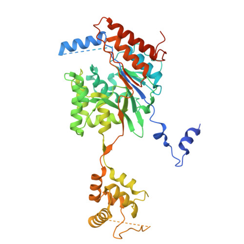
    Mutations that cause loss of function of GlcNAc-1-phosphotransferase (PTase) lead to the lysosomal storage disorder mucolipidosis II. PTase is the key enzyme of the mannose 6-phosphate (M6P) targeting system that is responsible for tagging lysosomal hydrolases with the M6P moiety for their delivery to the lysosome. We had previously generated a truncated hyperactive form of PTase termed S1S3 which was shown to notably increase the phosphorylation level of secreted lysosomal enzymes and enhance their uptake by cells. Here, we report the 3.4 Å cryo-EM structure of soluble S1S3 lacking both transmembrane domains and cytosolic tails. The structure reveals a high degree of conservation of the catalytic core to full-length PTase. In this dimeric structure, the EF-hand of one protomer is observed interacting with the conserved region four of the other. In addition, we present a high-quality EM 3D map of the UDP-GlcNAc bound form of the full-length soluble protein showing the key molecular interactions between the nucleotide sugar donor and side chain amino acids of the protein. Finally, although the domain organization of S1S3 is very similar to that of the Drosophila melanogaster (fruit fly) PTase homolog, we establish that the latter does not act on lysosomal hydrolases.


  • Organizational Affiliation
    • Department of Structural Biology, Van Andel Institute, Grand Rapids, Michigan, USA.

Macromolecules
Find similar proteins by:  (by identity cutoff)  |  3D Structure
Entity ID: 1
MoleculeChains Sequence LengthOrganismDetailsImage
highly active truncation of GlcNAc-1-phosphotransferase, S1S3,N-acetylglucosamine-1-phosphotransferase subunit beta
A, B
499Homo sapiensMutation(s): 0 
EC: 2.7.8.17
UniProt & NIH Common Fund Data Resources
Find proteins for Q3T906 (Homo sapiens)
Explore Q3T906 
Go to UniProtKB:  Q3T906
PHAROS:  Q3T906
GTEx:  ENSG00000111670 
Entity Groups  
Sequence Clusters30% Identity50% Identity70% Identity90% Identity95% Identity100% Identity
UniProt GroupQ3T906
Glycosylation
Glycosylation Sites: 2Go to GlyGen: Q3T906-1
Sequence Annotations
Expand
  • Reference Sequence
Experimental Data & Validation

Experimental Data

  • Method: ELECTRON MICROSCOPY
  • Resolution: 3.40 Å
  • Aggregation State: PARTICLE 
  • Reconstruction Method: SINGLE PARTICLE 
EM Software:
TaskSoftware PackageVersion
MODEL REFINEMENTPHENIX1.21.1_5286:

Structure Validation

View Full Validation Report


Entry History & Funding Information

Deposition Data

  • Released Date: 2025-02-26 
  • Deposition Author(s): Li, H., Li, H.

Funding OrganizationLocationGrant Number
National Institutes of Health/National Institute of Neurological Disorders and Stroke (NIH/NINDS)United StatesNS127292

Revision History  (Full details and data files)

  • Version 1.0: 2025-02-26
    Type: Initial release