


Interactom of cellular retinol binding protein 3.
Golczak, M.To be published.
Experimental Data Snapshot
Starting Model: experimental
View more details
Entity ID: 1 | |||||
|---|---|---|---|---|---|
| Molecule | Chains | Sequence Length | Organism | Details | Image |
| Retinol-binding protein 5 | 139 | Homo sapiens | Mutation(s): 0 Gene Names: RBP5 | 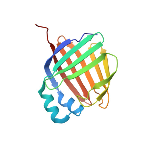 | |
UniProt & NIH Common Fund Data Resources | |||||
PHAROS: P82980 GTEx: ENSG00000139194 | |||||
Entity Groups | |||||
| Sequence Clusters | 30% Identity50% Identity70% Identity90% Identity95% Identity100% Identity | ||||
| UniProt Group | P82980 | ||||
Sequence AnnotationsExpand | |||||
Reference Sequence | |||||
| Ligands 2 Unique | |||||
|---|---|---|---|---|---|
| ID | Chains | Name / Formula / InChI Key | 2D Diagram | 3D Interactions | |
| A1ALJ (Subject of Investigation/LOI) Download:Ideal Coordinates CCD File | C [auth A] | 1-[11-(dipyrrometheneboron difluoride)undecanoyl]-rac-glycerol C27 H41 B F2 N2 O4 MYLJZUQNPWUJDF-QHCPKHFHSA-N |  | ||
| GOL Download:Ideal Coordinates CCD File | D [auth B] | GLYCEROL C3 H8 O3 PEDCQBHIVMGVHV-UHFFFAOYSA-N |  | ||
| Length ( Å ) | Angle ( ˚ ) |
|---|---|
| a = 41.213 | α = 90 |
| b = 46.257 | β = 92.1 |
| c = 71.53 | γ = 90 |
| Software Name | Purpose |
|---|---|
| PHENIX | refinement |
| XDS | data reduction |
| Aimless | data scaling |
| BALBES | phasing |
| Funding Organization | Location | Grant Number |
|---|---|---|
| National Institutes of Health/National Eye Institute (NIH/NEI) | United States | EY023948 |


