9B36 | pdb_00009b36

Open state of kainate receptor GluK2 in complex with agonist glutamate and positive allosteric modulator BPAM344 bound to two concanavalin A dimers. Composite map.

  • Classification: MEMBRANE PROTEIN
  • Organism(s): Rattus norvegicus, Canavalia ensiformis
  • Expression System: Homo sapiens
  • Mutation(s): No 
  • Membrane Protein: Yes  PDBTM

  • Deposited: 2024-03-18 Released: 2024-05-22 
  • Deposition Author(s): Nadezhdin, K.D., Gangwar, S.P., Sobolevsky, A.I.
  • Funding Organization(s): National Institutes of Health/National Institute of Neurological Disorders and Stroke (NIH/NINDS), National Institutes of Health/National Institute of Arthritis and Musculoskeletal and Skin Diseases (NIH/NIAMS), National Institutes of Health/National Cancer Institute (NIH/NCI)

Experimental Data Snapshot

  • Method: ELECTRON MICROSCOPY
  • Resolution: 4.29 Å
  • Aggregation State: PARTICLE 
  • Reconstruction Method: SINGLE PARTICLE 

wwPDB Validation 3D Report Full Report

Validation slider image for 9B36

This is version 1.3 of the entry. See complete history

Literature

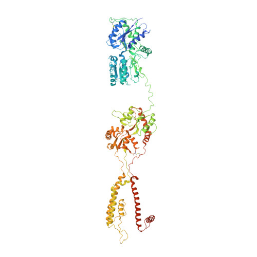
Kainate receptor channel opening and gating mechanism.

Gangwar, S.P.Yelshanskaya, M.V.Nadezhdin, K.D.Yen, L.Y.Newton, T.P.Aktolun, M.Kurnikova, M.G.Sobolevsky, A.I.

(2024) Nature 630: 762-768

  • DOI: https://doi.org/10.1038/s41586-024-07475-0
  • Primary Citation Related Structures: 
    9B33, 9B34, 9B35, 9B36, 9B37, 9B38, 9B39

  • PubMed Abstract: 

    Kainate receptors, a subclass of ionotropic glutamate receptors, are tetrameric ligand-gated ion channels that mediate excitatory neurotransmission 1-4 . Kainate receptors modulate neuronal circuits and synaptic plasticity during the development and function of the central nervous system and are implicated in various neurological and psychiatric diseases, including epilepsy, depression, schizophrenia, anxiety and autism 5-11 . Although structures of kainate receptor domains and subunit assemblies are available 12-18 , the mechanism of kainate receptor gating remains poorly understood. Here we present cryo-electron microscopy structures of the kainate receptor GluK2 in the presence of the agonist glutamate and the positive allosteric modulators lectin concanavalin A and BPAM344. Concanavalin A and BPAM344 inhibit kainate receptor desensitization and prolong activation by acting as a spacer between the amino-terminal and ligand-binding domains and a stabilizer of the ligand-binding domain dimer interface, respectively. Channel opening involves the kinking of all four pore-forming M3 helices. Our structures reveal the molecular basis of kainate receptor gating, which could guide the development of drugs for treatment of neurological disorders.


  • Organizational Affiliation
    • Department of Biochemistry and Molecular Biophysics, Columbia University, New York, NY, USA.

Macromolecule Content 

  • Total Structure Weight: 549.86 kDa 
  • Atom Count: 36,342 
  • Modeled Residue Count: 4,316 
  • Deposited Residue Count: 4,596 
  • Unique protein chains: 2

Macromolecules

Find similar proteins by:|  3D Structure
Entity ID: 1
MoleculeChains  Sequence LengthOrganismDetailsImage
Glutamate receptor ionotropic, kainate 2
A, B, C, D
912Rattus norvegicusMutation(s): 0 
Gene Names: Grik2Glur6
Membrane Entity: Yes 
UniProt
Find proteins for P42260 (Rattus norvegicus)
Explore P42260 
Go to UniProtKB:  P42260
Entity Groups
Sequence Clusters30% Identity50% Identity70% Identity90% Identity95% Identity100% Identity
UniProt GroupP42260
Glycosylation
Glycosylation Sites: 7Go to GlyGen: P42260-1
Sequence Annotations
Expand
Reference Sequence
Find similar proteins by:|  3D Structure
Entity ID: 2
MoleculeChains  Sequence LengthOrganismDetailsImage
Concanavalin A
E, F, G, H
237Canavalia ensiformisMutation(s): 0 
UniProt
Find proteins for C0HJY1 (Canavalia cathartica)
Explore C0HJY1 
Go to UniProtKB:  C0HJY1
Entity Groups
Sequence Clusters30% Identity50% Identity70% Identity90% Identity95% Identity100% Identity
UniProt GroupC0HJY1
Sequence Annotations
Expand
Reference Sequence

Oligosaccharides

Help  
Entity ID: 3
MoleculeChains Length2D Diagram GlycosylationD Interactions
beta-D-galactopyranose-(1-4)-2-acetamido-2-deoxy-beta-D-glucopyranose-(1-2)-alpha-D-mannopyranose-(1-6)-[alpha-D-mannopyranose-(1-3)]beta-D-mannopyranose-(1-4)-2-acetamido-2-deoxy-beta-D-glucopyranose-(1-4)-2-acetamido-2-deoxy-beta-D-glucopyranose
I, W
7N-Glycosylation
Glycosylation Resources
GlyTouCan: G28663KH
GlyCosmos: G28663KH
GlyGen: G28663KH
Entity ID: 4
MoleculeChains Length2D Diagram GlycosylationD Interactions
2-acetamido-2-deoxy-beta-D-glucopyranose-(1-4)-2-acetamido-2-deoxy-beta-D-glucopyranose
BA [auth b],
CA [auth c],
EA [auth e],
J,
JA [auth j],
BA [auth b],
CA [auth c],
EA [auth e],
J,
JA [auth j],
L,
N,
O,
Q,
V,
X,
Z
2N-Glycosylation
Glycosylation Resources
GlyTouCan: G42666HT
GlyCosmos: G42666HT
GlyGen: G42666HT
Entity ID: 5
MoleculeChains Length2D Diagram GlycosylationD Interactions
beta-D-galactopyranose-(1-4)-2-acetamido-2-deoxy-beta-D-glucopyranose-(1-2)-alpha-D-mannopyranose-(1-6)-[2-acetamido-2-deoxy-beta-D-glucopyranose-(1-2)-alpha-D-mannopyranose-(1-3)]beta-D-mannopyranose-(1-4)-2-acetamido-2-deoxy-beta-D-glucopyranose-(1-4)-2-acetamido-2-deoxy-beta-D-glucopyranose
K
8N-Glycosylation
Glycosylation Resources
GlyTouCan: G89993FE
GlyCosmos: G89993FE
GlyGen: G89993FE
Entity ID: 6
MoleculeChains Length2D Diagram GlycosylationD Interactions
beta-D-mannopyranose-(1-4)-2-acetamido-2-deoxy-beta-D-glucopyranose-(1-4)-2-acetamido-2-deoxy-beta-D-glucopyranoseAA [auth a],
GA [auth g],
M,
S,
T
3N-Glycosylation
Glycosylation Resources
GlyTouCan: G15407YE
GlyCosmos: G15407YE
GlyGen: G15407YE
Entity ID: 7
MoleculeChains Length2D Diagram GlycosylationD Interactions
beta-D-mannopyranose-(1-3)-beta-D-mannopyranose-(1-4)-2-acetamido-2-deoxy-beta-D-glucopyranose-(1-4)-2-acetamido-2-deoxy-beta-D-glucopyranoseDA [auth d],
P
4N-Glycosylation
Glycosylation Resources
GlyTouCan: G31886NL
GlyCosmos: G31886NL
GlyGen: G31886NL
Entity ID: 8
MoleculeChains Length2D Diagram GlycosylationD Interactions
2-acetamido-2-deoxy-beta-D-glucopyranose-(1-2)-alpha-D-mannopyranose-(1-3)-[2-acetamido-2-deoxy-beta-D-glucopyranose-(1-2)-alpha-D-mannopyranose-(1-6)]beta-D-mannopyranose-(1-4)-2-acetamido-2-deoxy-beta-D-glucopyranose-(1-4)-2-acetamido-2-deoxy-beta-D-glucopyranoseFA [auth f],
R,
Y
7N-Glycosylation
Glycosylation Resources
GlyTouCan: G39213VZ
GlyCosmos: G39213VZ
GlyGen: G39213VZ
Entity ID: 9
MoleculeChains Length2D Diagram GlycosylationD Interactions
alpha-D-mannopyranose-(1-6)-beta-D-mannopyranose-(1-4)-2-acetamido-2-deoxy-beta-D-glucopyranose-(1-4)-2-acetamido-2-deoxy-beta-D-glucopyranoseHA [auth h],
IA [auth i],
U
4N-Glycosylation
Glycosylation Resources
GlyTouCan: G22573RC
GlyCosmos: G22573RC
GlyGen: G22573RC

Small Molecules

Ligands 6 Unique
IDChains Name / Formula / InChI Key2D Diagram3D Interactions
POV

Query on POV


Download:Ideal Coordinates CCD File
AB [auth C]
DB [auth C]
EB [auth C]
FB [auth C]
JB [auth D]
AB [auth C],
DB [auth C],
EB [auth C],
FB [auth C],
JB [auth D],
MA [auth A],
MB [auth D],
NA [auth A],
NB [auth D],
OA [auth A],
TA [auth A],
UA [auth B],
XA [auth B],
YA [auth B]
(2S)-3-(hexadecanoyloxy)-2-[(9Z)-octadec-9-enoyloxy]propyl 2-(trimethylammonio)ethyl phosphate
C42 H82 N O8 P
WTJKGGKOPKCXLL-PFDVCBLKSA-N
CLR

Query on CLR


Download:Ideal Coordinates CCD File
GB [auth C]
HB [auth C]
IB [auth C]
PA [auth A]
QA [auth A]
GB [auth C],
HB [auth C],
IB [auth C],
PA [auth A],
QA [auth A],
RA [auth A],
SA [auth A],
ZA [auth B]
CHOLESTEROL
C27 H46 O
HVYWMOMLDIMFJA-DPAQBDIFSA-N
2J9
(Subject of Investigation/LOI)

Query on 2J9


Download:Ideal Coordinates CCD File
BB [auth C],
KA [auth A],
KB [auth D],
VA [auth B]
4-cyclopropyl-7-fluoro-3,4-dihydro-2H-1,2,4-benzothiadiazine 1,1-dioxide
C10 H11 F N2 O2 S
FLTMTBPCYAZIKM-UHFFFAOYSA-N
GLU
(Subject of Investigation/LOI)

Query on GLU


Download:Ideal Coordinates CCD File
CB [auth C],
LA [auth A],
LB [auth D],
WA [auth B]
GLUTAMIC ACID
C5 H9 N O4
WHUUTDBJXJRKMK-VKHMYHEASA-N
ZN

Query on ZN


Download:Ideal Coordinates CCD File
OB [auth E],
QB [auth F],
SB [auth G],
UB [auth H]
ZINC ION
Zn
PTFCDOFLOPIGGS-UHFFFAOYSA-N
CA

Query on CA


Download:Ideal Coordinates CCD File
PB [auth E],
RB [auth F],
TB [auth G],
VB [auth H]
CALCIUM ION
Ca
BHPQYMZQTOCNFJ-UHFFFAOYSA-N

Experimental Data & Validation

Experimental Data

  • Method: ELECTRON MICROSCOPY
  • Resolution: 4.29 Å
  • Aggregation State: PARTICLE 
  • Reconstruction Method: SINGLE PARTICLE 
EM Software:
TaskSoftware PackageVersion
MODEL REFINEMENTPHENIX1.18
RECONSTRUCTIONcryoSPARC4.4.1

Structure Validation

View Full Validation Report


Entry History 

& Funding Information

Deposition Data


Funding OrganizationLocationGrant Number
National Institutes of Health/National Institute of Neurological Disorders and Stroke (NIH/NINDS)United StatesR01 NS083660
National Institutes of Health/National Institute of Neurological Disorders and Stroke (NIH/NINDS)United StatesR01 NS107253
National Institutes of Health/National Institute of Arthritis and Musculoskeletal and Skin Diseases (NIH/NIAMS)United StatesR01 AR078814
National Institutes of Health/National Cancer Institute (NIH/NCI)United StatesR01 CA206573

Revision History  (Full details and data files)

  • Version 1.0: 2024-05-22
    Type: Initial release
  • Version 1.1: 2024-06-05
    Changes: Database references
  • Version 1.2: 2024-06-26
    Changes: Data collection, Database references
  • Version 1.3: 2024-11-13
    Changes: Data collection, Structure summary