9AZO | pdb_00009azo

Crystal structure of CHMS Dehydrogenase PmdC from Comamonas testosteroni bound to cofactor NADP


Experimental Data Snapshot

  • Method: X-RAY DIFFRACTION
  • Resolution: 2.34 Å
  • R-Value Free: 
    0.277 (Depositor), 0.277 (DCC) 
  • R-Value Work: 
    0.239 (Depositor), 0.239 (DCC) 
  • R-Value Observed: 
    0.240 (Depositor) 

Starting Model: in silico
View more details

wwPDB Validation 3D Report Full Report

Validation slider image for 9AZO

Ligand Structure Quality Assessment 


This is version 1.1 of the entry. See complete history

Literature

Characterization of lignin-degrading enzyme PmdC, which catalyzes a key step in the synthesis of polymer precursor 2-pyrone-4,6-dicarboxylic acid.

Rodrigues, A.V.Moriarty, N.W.Kakumanu, R.DeGiovanni, A.Pereira, J.H.Gin, J.W.Chen, Y.Baidoo, E.E.K.Petzold, C.J.Adams, P.D.

(2024) J Biological Chem 300: 107736-107736

  • DOI: https://doi.org/10.1016/j.jbc.2024.107736
  • Primary Citation Related Structures: 
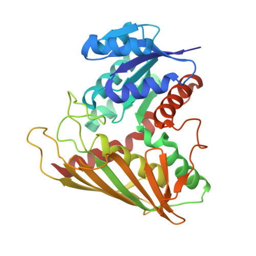
    9AZO

  • PubMed Abstract: 

    Pyrone-2,4-dicarboxylic acid (PDC) is a valuable polymer precursor that can be derived from the microbial degradation of lignin. The key enzyme in the microbial production of PDC is CHMS dehydrogenase, which acts on the substrate 4-carboxy-2-hydroxymuconate-6-semialdehyde (CHMS). We present the crystal structure of CHMS dehydrogenase (PmdC from Comamonas testosteroni) bound to the cofactor NADP, shedding light on its three-dimensional architecture, and revealing residues responsible for binding NADP. Using a combination of structural homology, molecular docking, and quantum chemistry calculations we have predicted the binding site of CHMS. Key histidine residues in a conserved sequence are identified as crucial for binding the hydroxyl group of CHMS and facilitating dehydrogenation with NADP. Mutating these histidine residues results in a loss of enzyme activity, leading to a proposed model for the enzyme's mechanism. These findings are expected to help guide efforts in protein and metabolic engineering to enhance PDC yields in biological routes to polymer feedstock synthesis.


  • Organizational Affiliation
    • Joint BioEnergy Institute, Emeryville, California, 94608, United States; Molecular Biophysics and Integrated Bioimaging, Lawrence Berkeley National Laboratory, Berkeley California 94720, United States. Electronic address: avrodrigues@lbl.gov.

Macromolecule Content 

  • Total Structure Weight: 216.64 kDa 
  • Atom Count: 15,371 
  • Modeled Residue Count: 1,890 
  • Deposited Residue Count: 1,914 
  • Unique protein chains: 1

Macromolecules

Find similar proteins by:|  3D Structure
Entity ID: 1
MoleculeChains  Sequence LengthOrganismDetailsImage
PmdC
A, B, C, D, E
A, B, C, D, E, F
319N/AMutation(s): 0 
EC: 1
UniProt
Find proteins for Q93PS4 (Comamonas testosteroni)
Explore Q93PS4 
Go to UniProtKB:  Q93PS4
Entity Groups
Sequence Clusters30% Identity50% Identity70% Identity90% Identity95% Identity100% Identity
UniProt GroupQ93PS4
Sequence Annotations
Expand
Reference Sequence

Small Molecules

Ligands 2 Unique
IDChains Name / Formula / InChI Key2D Diagram3D Interactions
NAP
(Subject of Investigation/LOI)

Query on NAP


Download:Ideal Coordinates CCD File
H [auth A]
J [auth B]
L [auth C]
N [auth D]
P [auth E]
H [auth A],
J [auth B],
L [auth C],
N [auth D],
P [auth E],
R [auth F]
NADP NICOTINAMIDE-ADENINE-DINUCLEOTIDE PHOSPHATE
C21 H28 N7 O17 P3
XJLXINKUBYWONI-NNYOXOHSSA-N
SO4

Query on SO4


Download:Ideal Coordinates CCD File
G [auth A]
I [auth B]
K [auth C]
M [auth D]
O [auth E]
G [auth A],
I [auth B],
K [auth C],
M [auth D],
O [auth E],
Q [auth F],
S [auth F]
SULFATE ION
O4 S
QAOWNCQODCNURD-UHFFFAOYSA-L

Experimental Data & Validation

Experimental Data

  • Method: X-RAY DIFFRACTION
  • Resolution: 2.34 Å
  • R-Value Free:  0.277 (Depositor), 0.277 (DCC) 
  • R-Value Work:  0.239 (Depositor), 0.239 (DCC) 
  • R-Value Observed: 0.240 (Depositor) 
Space Group: P 1 21 1
Unit Cell:
Length ( Å )Angle ( ˚ )
a = 79.151α = 90
b = 157.864β = 114.449
c = 95.011γ = 90
Software Package:
Software NamePurpose
PHENIXrefinement
PHENIXphasing

Structure Validation

View Full Validation Report


Ligand Structure Quality Assessment 


Entry History 

& Funding Information

Deposition Data


Funding OrganizationLocationGrant Number
Department of Energy (DOE, United States)United StatesDE-AC0205CH11231

Revision History  (Full details and data files)

  • Version 1.0: 2024-09-18
    Type: Initial release
  • Version 1.1: 2024-10-16
    Changes: Database references, Structure summary