8ZHA | pdb_00008zha

HIV-1 integrase core domain in complex with compound 15


Experimental Data Snapshot

  • Method: X-RAY DIFFRACTION
  • Resolution: 1.95 Å
  • R-Value Free: 
    0.211 (Depositor), 0.209 (DCC) 
  • R-Value Work: 
    0.190 (Depositor), 0.192 (DCC) 
  • R-Value Observed: 
    0.191 (Depositor) 

Starting Model: experimental
View more details

wwPDB Validation 3D Report Full Report

Validation slider image for 8ZHA

Ligand Structure Quality Assessment 


This is version 1.2 of the entry. See complete history

Literature

Design and synthesis of novel and potent allosteric HIV-1 integrase inhibitors with a spirocyclic moiety.

Adachi, K.Manabe, T.Yamasaki, T.Suma, A.Orita, T.Furuzono, T.Adachi, T.Ohata, Y.Akiyama, Y.Miyazaki, S.

(2024) Bioorg Med Chem Lett 110: 129864-129864

  • DOI: https://doi.org/10.1016/j.bmcl.2024.129864
  • Primary Citation Related Structures: 
    8ZH4, 8ZHA

  • PubMed Abstract: 

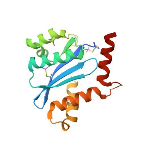
    We report herein the design and discovery of novel allosteric HIV-1 integrase inhibitors. Our design concept utilized the spirocyclic moiety to restrain the flexibility of the conformation of the lipophilic part of the inhibitor. Compound 5 showed antiviral activity by binding to the nuclear lens epithelium-derived growth factor (LEDGF/p75) binding site of HIV-1 integrase (IN). The introduction of a lipophilic amide substituent into the central benzene ring resulted in a significant increase in antiviral activity against HIV-1 WT X-ray crystallography of compound 15 in complex with the integrase revealed the presence of a hydrogen bond between the oxygen atom of the amide of compound 15 and the hydroxyl group of the T125 side chain. Chiral compound 17 showed high antiviral activity, good bioavailability, and low clearance in rats.


  • Organizational Affiliation
    • Chemical Research Laboratories, Central Pharmaceutical Research Institute, Japan Tobacco Inc., 1-1, Murasaki-cho, Takatsuki, Osaka 569-1125, Japan. Electronic address: kaoru.adachi@jt.com.

Macromolecule Content 

  • Total Structure Weight: 19.43 kDa 
  • Atom Count: 1,257 
  • Modeled Residue Count: 143 
  • Deposited Residue Count: 166 
  • Unique protein chains: 1

Macromolecules

Find similar proteins by:|  3D Structure
Entity ID: 1
MoleculeChains  Sequence LengthOrganismDetailsImage
Integrase166Human immunodeficiency virus type 1 (NEW YORK-5 ISOLATE)Mutation(s): 0 
Gene Names: gag-pol
EC: 2.7.7 (PDB Primary Data), 3.1 (PDB Primary Data)
UniProt
Find proteins for P12497 (Human immunodeficiency virus type 1 group M subtype B (isolate NY5))
Explore P12497 
Go to UniProtKB:  P12497
Entity Groups
Sequence Clusters30% Identity50% Identity70% Identity90% Identity95% Identity100% Identity
UniProt GroupP12497
Sequence Annotations
Expand
Reference Sequence

Small Molecules

Modified Residues  1 Unique
IDChains TypeFormula2D DiagramParent
CAF
Query on CAF
A
L-PEPTIDE LINKINGC5 H12 As N O3 SCYS

Experimental Data & Validation

Experimental Data

  • Method: X-RAY DIFFRACTION
  • Resolution: 1.95 Å
  • R-Value Free:  0.211 (Depositor), 0.209 (DCC) 
  • R-Value Work:  0.190 (Depositor), 0.192 (DCC) 
  • R-Value Observed: 0.191 (Depositor) 
Space Group: P 31 2 1
Unit Cell:
Length ( Å )Angle ( ˚ )
a = 72.74α = 90
b = 72.74β = 90
c = 66.45γ = 120
Software Package:
Software NamePurpose
PHENIXrefinement
DIALSdata reduction
Aimlessdata scaling
MOLREPphasing

Structure Validation

View Full Validation Report


Ligand Structure Quality Assessment 


Entry History 

& Funding Information

Deposition Data


Funding OrganizationLocationGrant Number
Not funded--

Revision History  (Full details and data files)

  • Version 1.0: 2024-07-10
    Type: Initial release
  • Version 1.1: 2024-07-24
    Changes: Database references
  • Version 1.2: 2024-10-23
    Changes: Structure summary