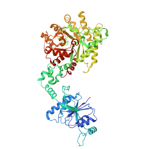
8Z40 | pdb_00008z40

The structure of type III CRISPR-associated deaminase apo form


Experimental Data Snapshot

  • Method: ELECTRON MICROSCOPY
  • Resolution: 3.26 Å
  • Aggregation State: PARTICLE 
  • Reconstruction Method: SINGLE PARTICLE 

wwPDB Validation 3D Report Full Report

Validation slider image for 8Z40

This is version 1.3 of the entry. See complete history. 

Literature

Antiviral signaling of a type III CRISPR-associated deaminase.

Li, Y., Li, Z., Yan, P., Hua, C., Kong, J., Wu, W., Cui, Y., Duan, Y., Li, S., Li, G., Ji, S., Chen, Y., Zhao, Y., Yang, P., Hu, C., Lu, M., Chen, M., Xiao, Y.

(2025) Science 387: eadr0393-eadr0393

  • DOI: https://doi.org/10.1126/science.adr0393
  • Primary Citation Related Structures: 
    8Z3K, 8Z3P, 8Z3R, 8Z40

  • PubMed Abstract: 

    Prokaryotes have evolved diverse defense strategies against viral infection, including foreign nucleic acid degradation by CRISPR-Cas systems and DNA and RNA synthesis inhibition through nucleotide pool depletion. Here, we report an antiviral mechanism of type III CRISPR-Cas-regulated adenosine triphosphate (ATP) depletion in which ATP is converted into inosine triphosphate (ITP) by CRISPR-Cas-associated adenosine deaminase (CAAD) upon activation by either cA 4 or cA 6 , followed by hydrolysis into inosine monophosphate (IMP) by Nudix hydrolase, ultimately resulting in cell growth arrest. The cryo-electron microscopy structures of CAAD in its apo and activated forms, together with biochemical evidence, revealed how cA 4 or cA 6 binds to the CRISPR-associated Rossmann fold (CARF) domain and abrogates CAAD autoinhibition, inducing substantial conformational changes that reshape the structure of CAAD and induce its deaminase activity. Our results reveal the mechanism of a CRISPR-Cas-regulated ATP depletion antiviral strategy.


  • Organizational Affiliation: 
    • Department of Pharmacology, School of Pharmacy, China Pharmaceutical University, Nanjing, China.

Macromolecule Content 

  • Total Structure Weight: 425.16 kDa 
  • Atom Count: 21,893 
  • Modeled Residue Count: 2,810 
  • Deposited Residue Count: 3,810 
  • Unique protein chains: 1

Macromolecules

Find similar proteins by:|  3D Structure
Entity ID: 1
MoleculeChains  Sequence LengthOrganismDetailsImage
Adenosine deaminase domain-containing protein
A, B, C, D, E
A, B, C, D, E, F
635Limisphaera ngatamarikiensisMutation(s): 0 
Gene Names: G4L39_03315
EC: 3.5.4.4
UniProt
Find proteins for A0A6M1RED6 (Limisphaera ngatamarikiensis)
Explore A0A6M1RED6 
Go to UniProtKB:  A0A6M1RED6
Entity Groups
Sequence Clusters30% Identity50% Identity70% Identity90% Identity95% Identity100% Identity
UniProt GroupA0A6M1RED6
Sequence Annotations
Expand
Reference Sequence

Experimental Data & Validation

Experimental Data

  • Method: ELECTRON MICROSCOPY
  • Resolution: 3.26 Å
  • Aggregation State: PARTICLE 
  • Reconstruction Method: SINGLE PARTICLE 
EM Software:
TaskSoftware PackageVersion
MODEL REFINEMENTPHENIX

Structure Validation

View Full Validation Report


Entry History 

& Funding Information

Deposition Data


Funding OrganizationLocationGrant Number
National Natural Science Foundation of China (NSFC)China--

Revision History  (Full details and data files)

  • Version 1.0: 2024-12-11
    Type: Initial release
  • Version 1.1: 2024-12-25
    Changes: Data collection, Database references
  • Version 1.2: 2025-03-05
    Changes: Data collection, Database references
  • Version 1.3: 2025-07-23
    Changes: Data collection, Database references