Mechanical activation opens a lipid-lined pore in OSCA ion channels.
Han, Y., Zhou, Z., Jin, R., Dai, F., Ge, Y., Ju, X., Ma, X., He, S., Yuan, L., Wang, Y., Yang, W., Yue, X., Chen, Z., Sun, Y., Corry, B., Cox, C.D., Zhang, Y.(2024) Nature 628: 910-918
- PubMed: 38570680 Search on PubMedSearch on PubMed Central
- DOI: https://doi.org/10.1038/s41586-024-07256-9
- Primary Citation Related Structures:
8XAJ, 8XNG, 8XRY, 8XS0, 8XS4, 8XS5, 8XVX, 8XVY, 8XVZ, 8XW0, 8XW1, 8XW2, 8XW3, 8XW4 - PubMed Abstract:
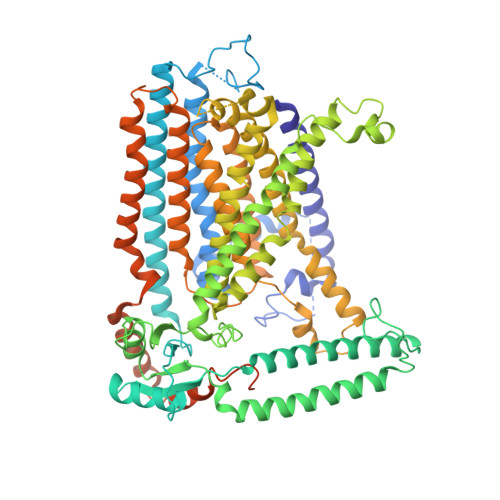
OSCA/TMEM63 channels are the largest known family of mechanosensitive channels 1-3 , playing critical roles in plant 4-7 and mammalian 8,9 mechanotransduction. Here we determined 44 cryogenic electron microscopy structures of OSCA/TMEM63 channels in different environments to investigate the molecular basis of OSCA/TMEM63 channel mechanosensitivity. In nanodiscs, we mimicked increased membrane tension and observed a dilated pore with membrane access in one of the OSCA1.2 subunits. In liposomes, we captured the fully open structure of OSCA1.2 in the inside-in orientation, in which the pore shows a large lateral opening to the membrane. Unusually for ion channels, structural, functional and computational evidence supports the existence of a 'proteo-lipidic pore' in which lipids act as a wall of the ion permeation pathway. In the less tension-sensitive homologue OSCA3.1, we identified an 'interlocking' lipid tightly bound in the central cleft, keeping the channel closed. Mutation of the lipid-coordinating residues induced OSCA3.1 activation, revealing a conserved open conformation of OSCA channels. Our structures provide a global picture of the OSCA channel gating cycle, uncover the importance of bound lipids and show that each subunit can open independently. This expands both our understanding of channel-mediated mechanotransduction and channel pore formation, with important mechanistic implications for the TMEM16 and TMC protein families.
- Interdisciplinary Research Center on Biology and Chemistry, Shanghai Institute of Organic Chemistry, Chinese Academy of Sciences, Shanghai, China.
Organizational Affiliation: