


The Crystal Structure of GSK3b from Biortus.
Wang, F., Cheng, W., Yuan, Z., Qi, J., Wu, B.To be published.
Experimental Data Snapshot
Starting Model: experimental
View more details
Entity ID: 1 | |||||
|---|---|---|---|---|---|
| Molecule | Chains | Sequence Length | Organism | Details | Image |
| Glycogen synthase kinase-3 beta | 421 | Trieres chinensis | Mutation(s): 0 Gene Names: GSK3B EC: 2.7.11.26 (PDB Primary Data), 2.7.11.1 (PDB Primary Data) | 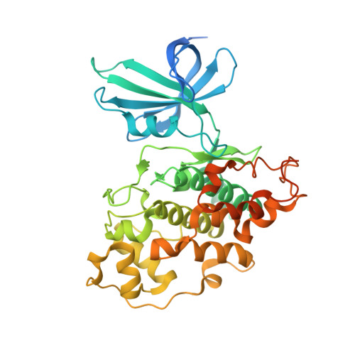 | |
UniProt & NIH Common Fund Data Resources | |||||
GTEx: ENSG00000082701 | |||||
Entity Groups | |||||
| Sequence Clusters | 30% Identity50% Identity70% Identity90% Identity95% Identity100% Identity | ||||
| UniProt Group | P49841 | ||||
Sequence AnnotationsExpand | |||||
Reference Sequence | |||||
| Ligands 4 Unique | |||||
|---|---|---|---|---|---|
| ID | Chains | Name / Formula / InChI Key | 2D Diagram | 3D Interactions | |
| ANP (Subject of Investigation/LOI) Download:Ideal Coordinates CCD File | C [auth A], P [auth B] | PHOSPHOAMINOPHOSPHONIC ACID-ADENYLATE ESTER C10 H17 N6 O12 P3 PVKSNHVPLWYQGJ-KQYNXXCUSA-N |  | ||
| PEG (Subject of Investigation/LOI) Download:Ideal Coordinates CCD File | AA [auth B], Y [auth B], Z [auth B] | DI(HYDROXYETHYL)ETHER C4 H10 O3 MTHSVFCYNBDYFN-UHFFFAOYSA-N |  | ||
| EDO (Subject of Investigation/LOI) Download:Ideal Coordinates CCD File | E [auth A] F [auth A] G [auth A] H [auth A] I [auth A] | 1,2-ETHANEDIOL C2 H6 O2 LYCAIKOWRPUZTN-UHFFFAOYSA-N |  | ||
| MG (Subject of Investigation/LOI) Download:Ideal Coordinates CCD File | D [auth A], Q [auth B] | MAGNESIUM ION Mg JLVVSXFLKOJNIY-UHFFFAOYSA-N |  | ||
| Length ( Å ) | Angle ( ˚ ) |
|---|---|
| a = 82.694 | α = 90 |
| b = 85.429 | β = 90 |
| c = 178.091 | γ = 90 |
| Software Name | Purpose |
|---|---|
| REFMAC | refinement |
| XDS | data reduction |
| Aimless | data scaling |
| PHASER | phasing |


