8XML | pdb_00008xml

Cryo-EM structure of the Apo CCR8-Gi complex


Experimental Data Snapshot

  • Method: ELECTRON MICROSCOPY
  • Resolution: 2.58 Å
  • Aggregation State: PARTICLE 
  • Reconstruction Method: SINGLE PARTICLE 

wwPDB Validation 3D Report Full Report

Validation slider image for 8XML

This is version 1.2 of the entry. See complete history

Literature

Cryo-EM Structure and Biochemical Analysis of the Human Chemokine Receptor CCR8.

Peng, Q.Jiang, H.Cheng, X.Wang, N.Zhou, S.Zhang, Y.Yang, T.Chen, Y.Zhang, W.Lv, S.Nan, W.Wang, J.Fan, G.H.Li, J.Zhang, J.

(2024) Biochemistry 63: 1892-1900

  • DOI: https://doi.org/10.1021/acs.biochem.4c00121
  • Primary Citation Related Structures: 
    8XML

  • PubMed Abstract: 

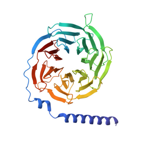
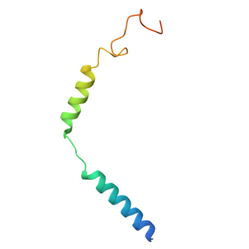
    The C-C motif chemokine receptor 8 (CCR8) is a class A G-protein-coupled receptor that has emerged as a promising therapeutic target in cancer and autoimmune diseases. In the present study, we solved the cryo-electron microscopy (cryo-EM) structure of the human CCR8-G i complex in the absence of a ligand at 2.58 Å. Structural analysis and comparison revealed that our apo CCR8 structure undergoes some conformational changes and is similar to that in the CCL1-CCR8 complex structure, indicating an active state. In addition, the key residues of CCR8 involved in the recognition of LMD-009, a potent nonpeptide agonist, were investigated by mutating CCR8 and testing the calcium flux induced by LMD-009-CCR8 interaction. Three mutants of CCR8, Y113 3.32 A, Y172 4.64 A, and E286 7.39 A, showed a dramatically decreased ability in mediating calcium mobilization, indicating their key interaction with LMD-009 and key roles in activation. These structural and biochemical analyses enrich molecular insights into the agonism and activation of CCR8 and will facilitate CCR8-targeted therapy.


  • Organizational Affiliation
    • The MOE Basic Research and Innovation Center for the Targeted Therapeutics of Solid Tumors, School of Basic Medical Sciences, Jiangxi Medical College, Nanchang University, Nanchang 330031, China.

Macromolecule Content 

  • Total Structure Weight: 167.08 kDa 
  • Atom Count: 8,245 
  • Modeled Residue Count: 1,071 
  • Deposited Residue Count: 1,493 
  • Unique protein chains: 5

Macromolecules

Find similar proteins by:|  3D Structure
Entity ID: 1
MoleculeChains  Sequence LengthOrganismDetailsImage
Soluble cytochrome b562,C-C chemokine receptor type 8A [auth R]461Escherichia coliHomo sapiens
This entity is chimeric
Mutation(s): 0 
Gene Names: cybCCCR8CKRL1CMKBR8CMKBRL2
Membrane Entity: Yes 
UniProt & NIH Common Fund Data Resources
Find proteins for P0ABE7 (Escherichia coli)
Explore P0ABE7 
Go to UniProtKB:  P0ABE7
Find proteins for P51685 (Homo sapiens)
Explore P51685 
Go to UniProtKB:  P51685
PHAROS:  P51685
GTEx:  ENSG00000179934 
Entity Groups
Sequence Clusters30% Identity50% Identity70% Identity90% Identity95% Identity100% Identity
UniProt GroupsP0ABE7P51685
Sequence Annotations
Expand
Reference Sequence
Find similar proteins by:|  3D Structure
Entity ID: 2
MoleculeChains  Sequence LengthOrganismDetailsImage
Guanine nucleotide-binding protein G(i) subunit alpha-1B [auth A]354Homo sapiensMutation(s): 5 
Gene Names: GNAI1
EC: 3.6.5
Membrane Entity: Yes 
UniProt & NIH Common Fund Data Resources
Find proteins for P63096 (Homo sapiens)
Explore P63096 
Go to UniProtKB:  P63096
PHAROS:  P63096
GTEx:  ENSG00000127955 
Entity Groups
Sequence Clusters30% Identity50% Identity70% Identity90% Identity95% Identity100% Identity
UniProt GroupP63096
Sequence Annotations
Expand
Reference Sequence
Find similar proteins by:|  3D Structure
Entity ID: 3
MoleculeChains  Sequence LengthOrganismDetailsImage
Guanine nucleotide-binding protein G(I)/G(S)/G(T) subunit beta-1C [auth B]340Homo sapiensMutation(s): 0 
Gene Names: GNB1
UniProt & NIH Common Fund Data Resources
Find proteins for P62873 (Homo sapiens)
Explore P62873 
Go to UniProtKB:  P62873
PHAROS:  P62873
GTEx:  ENSG00000078369 
Entity Groups
Sequence Clusters30% Identity50% Identity70% Identity90% Identity95% Identity100% Identity
UniProt GroupP62873
Sequence Annotations
Expand
Reference Sequence
Find similar proteins by:|  3D Structure
Entity ID: 4
MoleculeChains  Sequence LengthOrganismDetailsImage
Guanine nucleotide-binding protein G(I)/G(S)/G(O) subunit gamma-2D [auth G]71Homo sapiensMutation(s): 0 
Gene Names: GNG2
Membrane Entity: Yes 
UniProt & NIH Common Fund Data Resources
Find proteins for P59768 (Homo sapiens)
Explore P59768 
Go to UniProtKB:  P59768
PHAROS:  P59768
GTEx:  ENSG00000186469 
Entity Groups
Sequence Clusters30% Identity50% Identity70% Identity90% Identity95% Identity100% Identity
UniProt GroupP59768
Sequence Annotations
Expand
Reference Sequence
Find similar proteins by:|  3D Structure
Entity ID: 5
MoleculeChains  Sequence LengthOrganismDetailsImage
scFv16E [auth S]267Homo sapiensMutation(s): 0 

Experimental Data & Validation

Experimental Data

  • Method: ELECTRON MICROSCOPY
  • Resolution: 2.58 Å
  • Aggregation State: PARTICLE 
  • Reconstruction Method: SINGLE PARTICLE 
EM Software:
TaskSoftware PackageVersion
MODEL REFINEMENTPHENIX

Structure Validation

View Full Validation Report


Entry History 

& Funding Information

Deposition Data


Funding OrganizationLocationGrant Number
National Natural Science Foundation of China (NSFC)China--

Revision History  (Full details and data files)

  • Version 1.0: 2025-01-01
    Type: Initial release
  • Version 1.1: 2025-07-02
    Changes: Data collection
  • Version 1.2: 2026-01-14
    Changes: Data collection, Database references